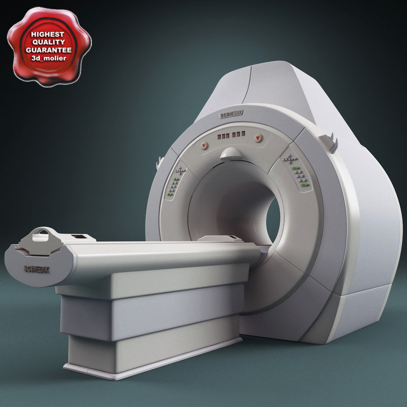
3D mri scanner
The Benefits of 3D MRI Prostate Mapping
3D MRI Prostate mapping is a new procedure which offers a number of improvements compared to the traditional TRUS (transrectal ultrasound) biopsy for people who have prostate cancer. TRUS guided biopsies have been the go-to way of diagnosing prostate malignancy for the last 20 years, but this procedure is quite invasive, and it is not very precise.
There is always the risk that an ultrasound will miss smaller tumors, because the soft tissues show up in low resolution on the scan. In addition, the grid approach used by the ultrasound will target the prostate gland’s peripheral aspects, and this means that they can miss 30-40 percent of the cancer – especially if it is in the anterior, midline or apex. There are also some risks associated with the number of needles used for this type of scan. The more needles that are used, the more accurate the scan, but also the greater the risk of infection and sexual or urinary side effects.
Why 3D MRI Scans Are Better
3D MRI prostate scans use Multi-Parametric MRI. There is no need to use an endorectal coil for the scan (unlike with older versions of the MRI technology), and the scan is far higher definition than an ultrasound. The procedure is non-invasive, and it allows for a detailed analysis of the entire prostate gland. This means that doctors can get a clear idea of how many tumors a patient has, and how aggressive the cancer is – potentially eliminating the need for a biopsy.
Because the 3D MRI scan is able to identify smaller tumors, it means that you get a more accurate diagnosis. In addition, you will be able to enjoy better treatment if you do need a biopsy, since the biopsy can be focused from the high resolution MRI.
The biopsy will target exactly the right tissue, so it is less invasive and healthy tissue will not be sampled. Side effects are rarer, and the risk of infections is reduced. The MRI biopsy will target only the tissue that is highly likely to contain cancer.
Unfortunately, prostate cancer cannot be diagnosed by MRI alone. The MRI can give an idea of blood flow, tissue density and other things which are indicators of a risk of cancer. There are certain characteristics of cancerous tissues which can be used to give doctors an idea of whether a tissue might be cancerous, but the tissue must be examined more closely to determine whether or not it is cancerous with more accuracy.
MRI-Guided biopsies help to ensure that the treatment you get matches the severity of the condition, and saves you from potential infections and long-lasting side effects. It reduces the risk of smaller cancerous areas (which are low risk, but could spread if the cells are aggressive), being missed.
When it comes to your health, it makes sense to not take any chances, and to arm yourself and your team of specialists with as much information as possible. Why use a 20-year-old technology, or the more invasive 1.5T MRI, which can be incredibly uncomfortable and may produce a distorted image because of the pressure of the coil on the prostate gland, when you can use the latest technologies and get a treatment which is accurate, produces fast results, and does so with minimum impact on your comfort.
Call your health insurance company today and ask them about 3D MRI Prostate scans. If you are at risk of prostate cancer or have been referred for diagnosis, this could be the perfect option for you.
More details are available on our page – Prostate Map Imaging
Magnetic Resonance Imaging (MRI)
- What is MRI?
- How does MRI work?
- What is MRI used for?
- Are there risks?
- What are examples of NIBIB-funded projects in MRI?
What is MRI?
Magnetic Resonance Imaging (MRI) is a non-invasive imaging technology that produces three dimensional detailed anatomical images. It is often used for disease detection, diagnosis, and treatment monitoring. It is based on sophisticated technology that excites and detects the change in the direction of the rotational axis of protons found in the water that makes up living tissues.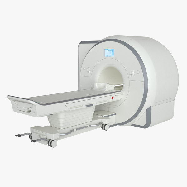
How does MRI work?
MRI of a kneeMRIs employ powerful magnets which produce a strong magnetic field that forces protons in the body to align with that field. When a radiofrequency current is then pulsed through the patient, the protons are stimulated, and spin out of equilibrium, straining against the pull of the magnetic field. When the radiofrequency field is turned off, the MRI sensors are able to detect the energy released as the protons realign with the magnetic field. The time it takes for the protons to realign with the magnetic field, as well as the amount of energy released, changes depending on the environment and the chemical nature of the molecules. Physicians are able to tell the difference between various types of tissues based on these magnetic properties.
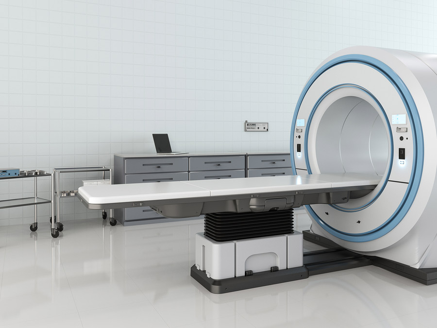
To obtain an MRI image, a patient is placed inside a large magnet and must remain very still during the imaging process in order not to blur the image. Contrast agents (often containing the element Gadolinium) may be given to a patient intravenously before or during the MRI to increase the speed at which protons realign with the magnetic field. The faster the protons realign, the brighter the image.
What is MRI used for?
MRI scanners are particularly well suited to image the non-bony parts or soft tissues of the body. They differ from computed tomography (CT), in that they do not use the damaging ionizing radiation of x-rays. The brain, spinal cord and nerves, as well as muscles, ligaments, and tendons are seen much more clearly with MRI than with regular x-rays and CT; for this reason MRI is often used to image knee and shoulder injuries.
In the brain, MRI can differentiate between white matter and grey matter and can also be used to diagnose aneurysms and tumors. Because MRI does not use x-rays or other radiation, it is the imaging modality of choice when frequent imaging is required for diagnosis or therapy, especially in the brain.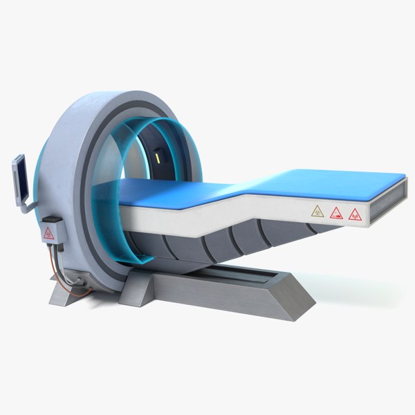 However, MRI is more expensive than x-ray imaging or CT scanning.
One kind of specialized MRI is functional Magnetic Resonance Imaging (fMRI.) This is used to observe brain structures and determine which areas of the brain “activate” (consume more oxygen) during various cognitive tasks. It is used to advance the understanding of brain organization and offers a potential new standard for assessing neurological status and neurosurgical risk.
Are there risks?
Although MRI does not emit the ionizing radiation that is found in x-ray and CT imaging, it does employ a strong magnetic field. The magnetic field extends beyond the machine and exerts very powerful forces on objects of iron, some steels, and other magnetizable objects; it is strong enough to fling a wheelchair across the room. Patients should notify their physicians of any form of medical or implant prior to an MR scan.
When having an MRI scan, the following should be taken into consideration:
- People with implants, particularly those containing iron, — pacemakers, vagus nerve stimulators, implantable cardioverter- defibrillators, loop recorders, insulin pumps, cochlear implants, deep brain stimulators, and capsules from capsule endoscopy should not enter an MRI machine.

- Noise—loud noise commonly referred to as clicking and beeping, as well as sound intensity up to 120 decibels in certain MR scanners, may require special ear protection.
- Nerve Stimulation—a twitching sensation sometimes results from the rapidly switched fields in the MRI.
- Contrast agents—patients with severe renal failure who require dialysis may risk a rare but serious illness called nephrogenic systemic fibrosis that may be linked to the use of certain gadolinium-containing agents, such as gadodiamide and others. Although a causal link has not been established, current guidelines in the United States recommend that dialysis patients should only receive gadolinium agents when essential, and that dialysis should be performed as soon as possible after the scan to remove the agent from the body promptly.
- Pregnancy—while no effects have been demonstrated on the fetus, it is recommended that MRI scans be avoided as a precaution especially in the first trimester of pregnancy when the fetus’ organs are being formed and contrast agents, if used, could enter the fetal bloodstream.
- Claustrophobia—people with even mild claustrophobia may find it difficult to tolerate long scan times inside the machine. Familiarization with the machine and process, as well as visualization techniques, sedation, and anesthesia provide patients with mechanisms to overcome their discomfort. Additional coping mechanisms include listening to music or watching a video or movie, closing or covering the eyes, and holding a panic button. The open MRI is a machine that is open on the sides rather than a tube closed at one end, so it does not fully surround the patient. It was developed to accommodate the needs of patients who are uncomfortable with the narrow tunnel and noises of the traditional MRI and for patients whose size or weight make the traditional MRI impractical. Newer open MRI technology provides high quality images for many but not all types of examinations.
What are examples of NIBIB-funded projects in MRI?
Replacing Biopsies with Sound
Chronic liver disease and cirrhosis affect more than 5.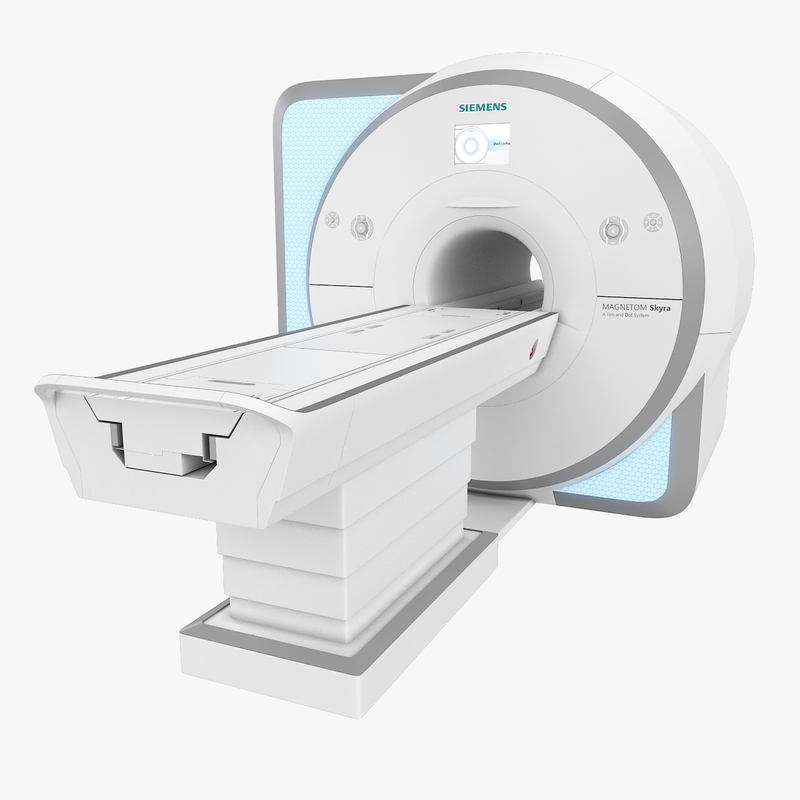 5 million people in the United States. NIBIB-funded researchers have developed a method to turn sound waves into images of the liver, which provides a new non-invasive, pain-free approach to find tumors or tissue damaged by liver disease. The Magnetic Resonance Elastography (MRE) device is placed over the liver of the patient before he enters the MRI machine. It then pulses sound waves through the liver, which the MRI is able to detect and use to determine the density and health of the liver tissue. This technique is safer and more comfortable for the patient as well as being less expensive than a traditional biopsy. Since MRE is able to recognize very slight differences in tissue density, there is the potential that it could also be used to detect cancer.
New MRI just for Kids
MRI is potentially one of the best imaging modalities for children since unlike CT, it does not have any ionizing radiation that could potentially be harmful. However, one of the most difficult challenges that MRI technicians face is obtaining a clear image, especially when the patient is a child or has some kind of ailment that prevents them from staying still for extended periods of time.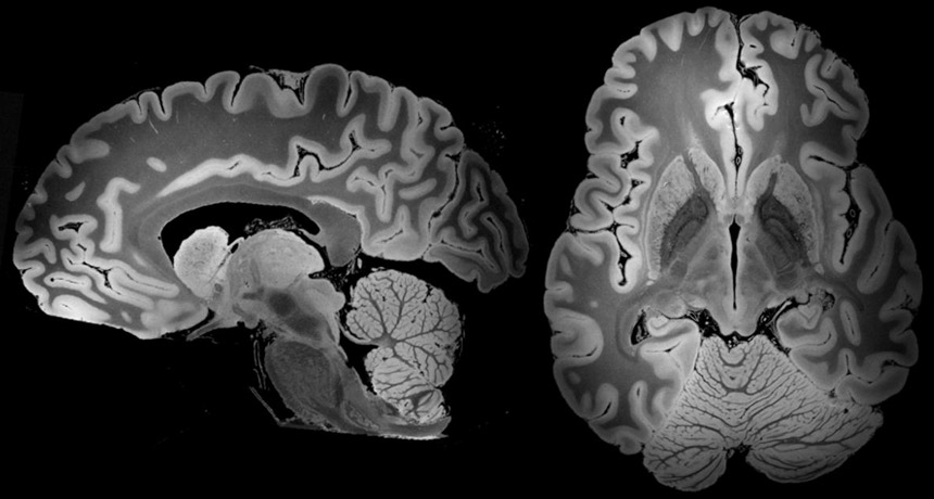 As a result, many young children require anesthesia, which increases the health risk for the patient. NIBIB is funding research that is attempting to develop a robust pediatric body MRI. By creating a pediatric coil made specifically for smaller bodies, the image can be rendered more clearly and quickly and will demand less MR operator skill. This will make MRIs cheaper, safer, and more available to children. The faster imaging and motion compensation could also potentially benefit adult patients as well.
Another NIBIB-funded researcher is trying to solve this problem from a different angle. He is developing a motion correction system that could greatly improve image quality for MR exams. Researchers are developing an optical tracking system that would be able to match and adapt the MRI pulses to changes in the patient’s pose in real time. This improvement could reduce cost (since less repeat MR exams will have to take place due to poor quality) as well as make MRI a viable option for many patients who are unable to remain still for the exam and reduce the amount of anesthesia used for MR exams.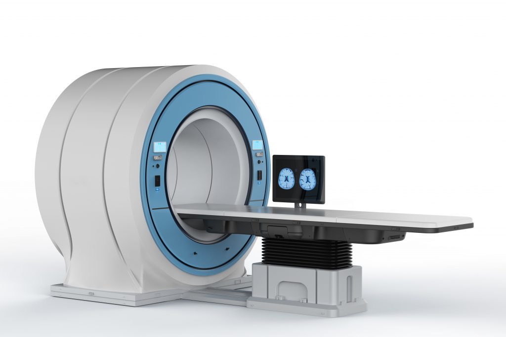
Determining the aggressiveness of a tumor
Traditional MRI, unlike PET or SPECT, cannot measure metabolic rates. However, researchers funded by NIBIB have discovered a way to inject specialized compounds (hyperpolarized carbon 13) into prostate cancer patients to measure the metabolic rate of a tumor. This information can provide a fast and accurate picture of the tumor’s aggressiveness. Monitoring disease progression can improve risk prediction, which is critical for prostate cancer patients who often adopt a wait and watch approach.
Thumbnail
mrithumbnail2_0.jpg
Smartphone, 3D printer, MRI scanner: interesting facts about modern technologies
Ekaterina Ushakhina
Did you know that sneakers, blood vessels and even bones can be 3D printed? Have you thought about how GPS navigation works on a smartphone? How does an airplane take off? How are a car, a vacuum cleaner, and a toaster arranged?
New technologies have changed all spheres of life - from transport to communications, from medicine to agriculture.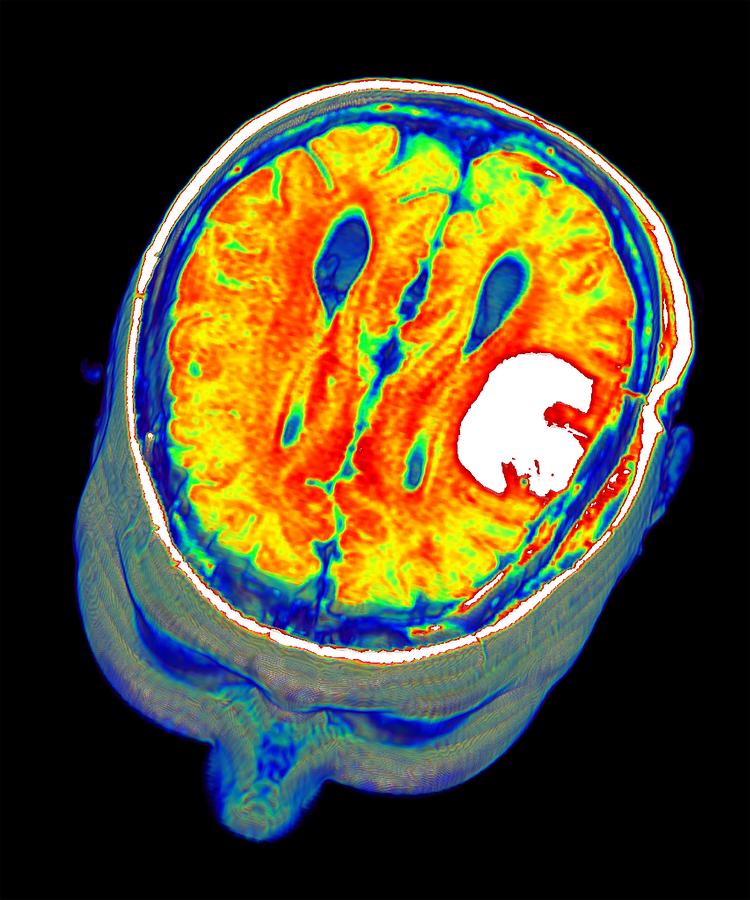 They affect what we eat, what we wear, how we move, and even how we communicate with friends. We are more and more dependent on technology. nine0004
How Technology Works is a collection of simple explanations, visual illustrations and interesting facts. They will help to understand the modern world and master the technologies that surround us.
How Technology Works
From Computing to Medicine
The new book is part of the DK Encyclopedia series of world-renowned encyclopedias. She vividly, in detail and entertainingly talks about the device of a variety of things that surround us: from a zipper to virtual reality and an MRI scanner. It reveals in great detail the topic of gadgets that simplify our lives: here children will see how a smartphone, smart watch, e-book and much more work. nine0004
The book has 9 large sections: from agriculture and energy to computer and medical technology.
The first chapter explains the principles behind many devices, from basic mechanics to electricity and digital technology.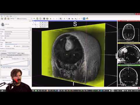 In the chapters that follow, devices are grouped by application, such as home, transport, computer.
The book not only describes the arrangement of everyday things and more complex mechanisms, but also tells about future technologies that will help save the environment: solar energy, electric and hybrid cars, and much more. nine0004
Navigation
Each spread here is a separate topic, which is revealed through beautiful diagrams and graphs. The book contains a sea of interesting and useful information, and convenient navigation (cross-references) allows you to move between chapters non-linearly in search of relationships.
Detailed instructions are supported by simple and original graphics that take the devices apart and show how they work.
On each spread you will find a detailed description of this or that invention, an interesting picture that will explain step by step how this or that object works. As well as a section that talks about unusual ways to use the device and curious facts (highlighted in large print). nine0004
3D printed blood vessels and bones
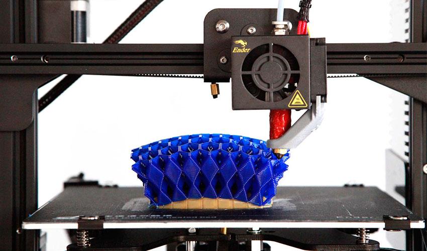
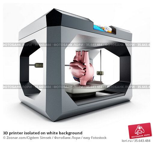
This is how the book talks about modern technology - 3D printing :
Conventional printing is applying a layer of ink to paper. A 3D printer works on the same principle, only it has to apply many layers to get a three-dimensional object. Thermoplastic plastic and other materials are used instead of ink. 3D printed products are not as perfect as industrial counterparts, but this method is sometimes faster and cheaper. nine0004
3D printing technology is recent and not yet used in mass production of consumer goods. In this way, specialized or piece goods are most often made.
3D printing technology can save us from having to transport some items over long distances.
In 2014, an astronaut on the International Space Station 3D printed a wrench using a file sent from Earth.
nine0054Here are some more amazing ways to use your 3D printer:
Artificial blood vessels.
Scientists create 3D-printed blood vessels containing living cells. Successful experiments on mice give hope that in the future such vessels can be introduced into the human body to replace damaged ones.
Sports shoes. Several athletic shoe companies produce 3D printed sneakers. Production is carried out on a limited scale, but some athletes have already been seen wearing such shoes at international competitions. nine0004
Artificial bones. For patients who have lost a piece of bone (for example, in the treatment of cancer), special prostheses made of titanium or synthetic material are created and implanted in the desired area.
Dentures. 3D-printed prosthetic limbs could be lighter than traditional ones. Their production is much cheaper, and such a prosthesis is easier to adapt to a particular patient.
Musical instruments. As an experiment, a variety of musical instruments were printed on a 3D printer: guitars, flutes, violins and other wind and string instruments.
Some of them went on sale. nine0004
Interesting facts
On the pages of the book you will find such amazing facts that they will amaze not only children, but also adults and, probably, even scientists!
They are short, beautifully designed and stand out from the rest of the text. Here are just a few of them.
— Since the first IVF procedure in 1978, more than 8 million test-tube babies have been born worldwide.
- The height of the world's first microwave oven is 1.7 meters.
— There are over 3 billion desktops and laptops in the world. nine0004
- 90 cm - the length of the keys that open the bomb-proof door to the Bank of England gold vault.
- There are more than 1 billion bicycles in the world, and their number is increasing by 100 million every year.
- 500 billion plastic bags are used daily in the world.
- One excavator replaces the labor of 174 people.
Modern technologies make our life easier and more interesting, help us achieve things that people of the past could not even dream of.
Do you want to know how modern things and technologies work?
Answers in How Technology Works.
GE Discovery MR450 1.5T MRI
The Discovery MR450 MR scanner brings the innovative technology of the Discovery MR750 scanner to the 1.5T world and delivers exceptional image quality. The tomograph provides at the same time convenience of the patient, productivity of work, excellent quality of the image. With this MRI system, the company's engineers managed to combine the necessary amount of movement with amazingly high image quality without compromise. nine0004
Good value for money makes it the ideal choice for power users looking for a versatile system for intensive use. The power of its magnet - 1.5 T - is used in the industry most often. And the diameter of the tunnel and the size of the field of view allow the use of this scanner in most patients who are indicated for MR imaging.

Scanner control and easy access to ECG and peripheral pulse synchronization leads and IV tubing are possible from either side of the patient table. Buttons corresponding to the next logical operation in the exam procedure are highlighted, making it easy to set up patient data. Patient data setup can be completed in 30 seconds with two simple steps. nine0004
A comprehensive suite of clinical applications, including IDEAL, LAVA-Flex, VIBRANT-Flex and Cube PIs, allows more patients to be examined by reducing the number of scans, increasing efficiency and helping clinics meet schedules more accurately.
Patients with chronic liver diseases such as fibrosis and cirrhosis often require regular follow-up with a gastroenterologist. This may require invasive procedures, which do not always provide complete information about the state of the liver. nine0151 The
Cube IP is a 3D isotropic resolution imaging technique - imaging is done once, with the ability to reformat images in any projection with excellent resolution. TheThe Cube UI uses a unique and innovative ARC accelerated data acquisition algorithm that reduces 3D rendering time and reduces voxel sizes to improve the quality of reformatted projections;
SWAN IP is a unique 3D T2*-weighted imaging technique with the ability to clearly highlight small vessels and diagnose microscopic bleeding. Like any 3D data acquisition application, the SWAN IP benefits from the increased signal-to-noise ratio provided by OpTix optical RF transmission technology; nine0151 The PROPELLER HD DWI suppresses magnetic susceptibility artifacts at the air/tissue interface that typically appear on temporal lobe images using standard echoplanar tomography techniques. This PI also almost completely suppresses artifacts from dental or surgical implants; PROPELLER HD delivers high-resolution, motion-insensitive axial, coronal, or sagittal images; nine0151 The Inhance 3D Velocity IP is a contrast-free technique designed to obtain angiographic images of the brain and renal arteries with excellent background suppression and short scan times. TheThis technique allows you to completely scan the entire neurovascular system in 5-6 minutes;
3D MERGE imager is designed to generate images of the musculoskeletal system and spine with increased contrast and signal-to-noise ratio. It also benefits from the improved signal-to-noise ratio provided by the OpTix technology; nine0151 The new MR450w wide-bore magnet delivers unrivaled image quality of off-center areas with uniform fat suppression; TheIDEAL PI is a reliable and high resolution alternative to the FSE Fat Sat or STIR PI for T2-weighted fat-saturation breast imaging and also suppresses surgical clip artefacts from surgery; nine0151 The Discovery MR450 creates stunning images of the breast using diffusion weighted imaging. For this purpose, a high-density phased coil for breast examinations or an integrated coil for examinations of the body, characterized by an increased imaging area, can be used; GE's unique VIBRANT technology creates up to 4 high-resolution, high-contrast breast images in a single short scan, and virtually eliminates fat suppression problems, even with large, irregularly shaped fields of view. 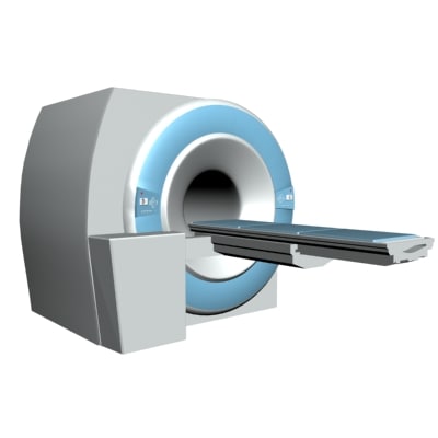
Learn more