3D printing knee cartilage
Scientists 3D bioprint articular cartilage protheses from stem cells
0Shares
The 2021 3D Printing Industry Awards shortlists are open for voting, have your say now.
Scientists from the Nakayama Lab at Saga University and Kyoto University in Japan have fabricated 3D printed cartilage constructs from stem cells which could potentially be used to repair large cartilage defects in patients.
Using human induced pluripotent stem cells (iPSCs), the researchers created the cartilage structures using a scaffold-free Kenzan bioprinting process, where the only support structure of the bioink is a microneedle matrix.
The technique reportedly enabled the scientists to overcome the limitations of scaffold-based tissue engineering methods for creating cartilage-like structures, such as poor cytocompatibility and toxicity, while solving the problem of bacterial infection and some risks associated with replacement surgery that conventional joint prostheses have.
The aim of the study is to treat conditions such as arthritis in athletes who do not want to undergo treatment with artificial joints made of metal and plastic.
Schematic representation of the 3D printing process. Image via Biofabrication.Improving arthritis treatment
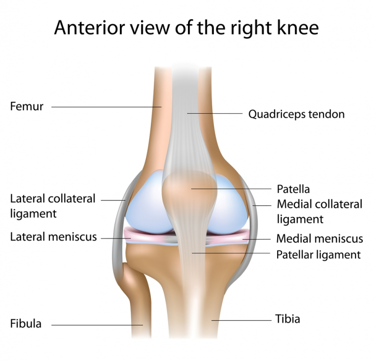
Articular cartilage is a load-bearing connective tissue that covers the ends of bones to prevent friction and protect joints. Cartilage damage can occur as a result of disease, trauma, and degenerative conditions such as osteoarthritis, one of the leading causes of joint pain and immobility.
Over 300 million people worldwide suffer from osteoarthritis, causing a huge clinical and economic burden. Current surgical treatments, such as microfractures and implants, typically only focus on focal defects and do not generate long-lasting functional cartilage. Total joint replacement is also the only treatment for end-stage osteoarthritis, with implants lacking durability over extended periods of time and potentially leading to bone shortening and other complications.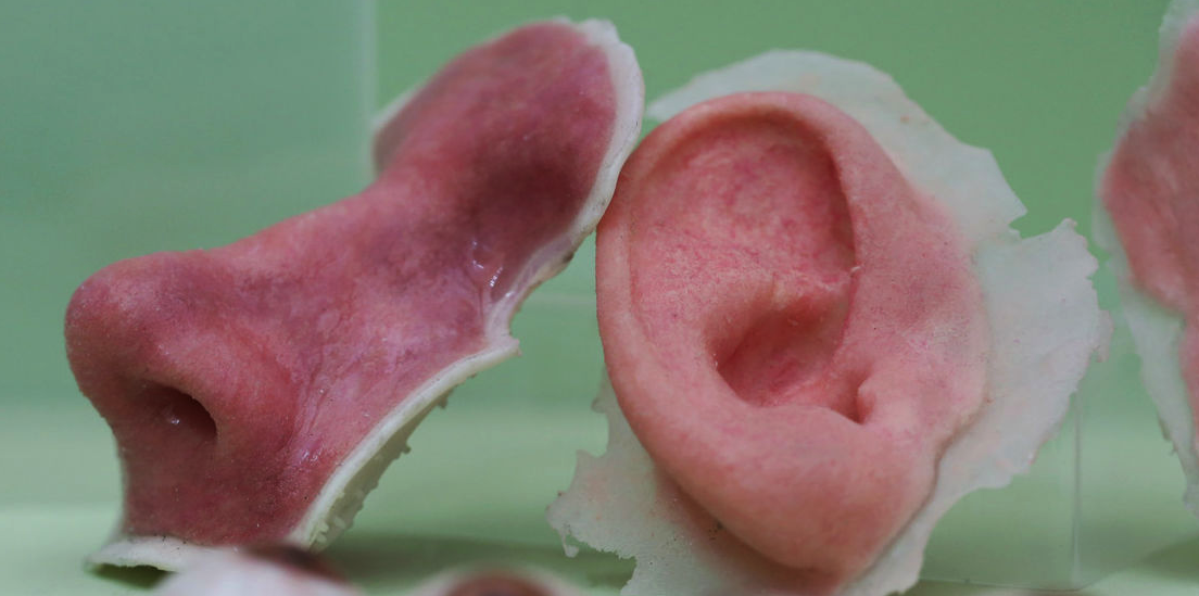
Tissue engineering is an alternative to total joint replacement for cartilage repair and joint preservation, however the method is limited by a lack of functional cell types and biomaterials. Chondrocytes and mesenchymal stem cells (MSCs) are currently considered the most promising sources of cells for regeneration therapy, although they can be hampered by donor age, density, activity, cell phenotype and other factors.
iPSCs are an alternative cell source that show promise in overcoming some of these limitations, due to their self-renewal potential to provide an almost unlimited number of cells with pluripotent capabilities – the ability to differentiate into many different cell types. As such, iPSCs were chosen as the basis of the scientists’ cartilage structures.
Bio-3D printer design data and images of the L-shaped construct removed from the Kenzan 18 days after printing. Image via Biofabrication.3D printing cartilage
Present tissue engineering technologies for cartilage regeneration can largely be separated into scaffold-based and scaffold-free methods, with 3D printing having been leveraged for multiple research projects in this area in the past.
For instance, scientists from the Chalmers University of Technology have previously demonstrated cartilage tissue engineering to treat osteoarthritis using 3D bioprinting, and microbiologists at Central Queensland University have explored a combination of crocodile protein and 3D bioprinting to repair joint damage. Elsewhere, Penn State researchers have developed a novel 3D printing method to create cartilage tissue building blocks with micropores that allow nutrient and oxygen diffusion.
More recently, researchers at the University of Alberta have developed a method of 3D bioprinting customized nasal cartilage for cancer patients living with post-operative facial disfiguration, and Swansea University is 3D printing cartilage tissue scaffolds for scar-free ear and nose transplants in partnership with the Scar Free Foundation.
One high-profile initiative that is currently making headway in this area is the EU-backed TRIANKLE project, which is seeking to develop 3D bioprinted cell-restoring ankle implants. Last year, the project received the backing of Spanish football club FC Barcelona’s R&D lab, which has agreed to test the collagen-based cell graft transplants on its amateur squad with a view to eventually prepare them for future commercial release.
3D printing cartilage protheses with iPSCs
The aim of the Japanese scientists’ current study was to leverage a 3D bioprinter to engineer a scaffold-free cartilage structure of anatomical size and shape. A Regenova Bio-3D printer from Cyfuse Biomedical was used for the study. The Regenova is a novel robotic system that facilitates the fabrication of 3D cellular structures by placing cellular spheroids in fine needle arrays, the Kenzan method, according to pre-designed 3D data.
The printer leveraged a 9 x 9 square Kenzan array consisting of 81 needles, a 26 x 26 square Kenzan array of 676 needles, and a 7 mm diameter round-type Kenzan array of 36 needles in a single row, in order to 3D print various sizes of cartilage protheses.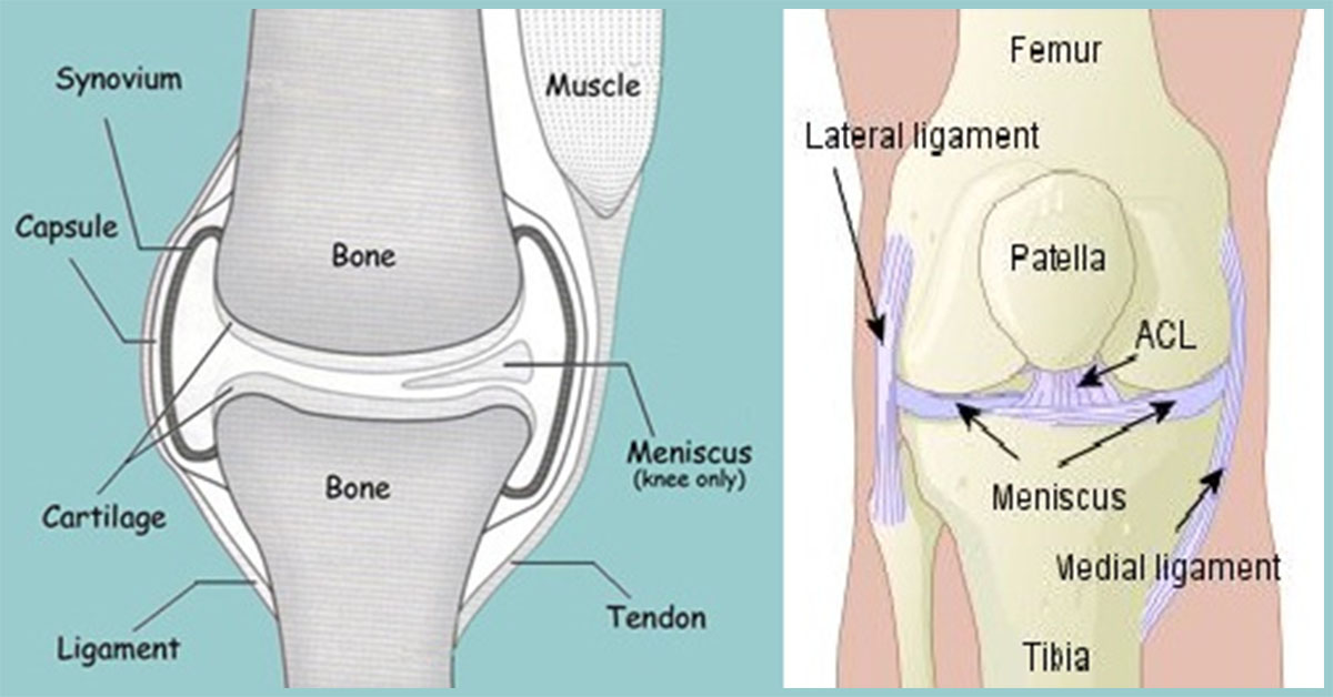
First, iPSCs were differentiated toward chondrocytes – metabolically active cells for cartilage formation – through MSC induction to form highly chondrogenic and potent iNCMSCs. The iNCMSCs were then optimized for the bioprinting process, after which a minimum maturation period was determined. Then, the various types of Kenzan listed above were deployed to fabricate larger cartilage constructs that were specifically shaped for irregular defects.
The first construct, which confirmed the method’s potential to sustain shape, was printed in an L-shape with a 90-degree angle. After removal from the Kenzan, the construct was able to maintain the applied design and withstand mechanical manipulation regardless of its 2 cm square total area.
Applying the 7 mm round Kenzan, the scientists were able to increase the construct size up to 6 cm squared in a half-pipe-shaped structure which could be utilized to treat a wide, curved area such as the patellar surface.
Bio-3D printer design data and images of the articular surface-shape construct.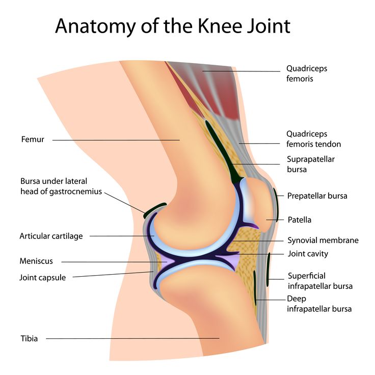 Image via Biofabrication.
Through their approach, the scientists were able to successfully 3D print scaffold-free, self-sustainable cartilage constructs of a suitable size and shape for repairing larger cartilage defects. The constructs were also found to exhibit a strength almost equal to that of normal cartilage.
The durability of the scientists’ cell-based joint prosthesis will continue to be assessed in future studies, and it is hoped that it will solve the problem of bacterial infection and some risks associated with replacement surgery that conventional joint prostheses have.
However, they did note that while using iPSC-derived cells as a source material has many benefits, it remains extremely expensive. Therefore, they state that further optimization and improvement of the technology will be crucial before they can conduct even more time and money-consuming studies in vitro.
Further information on the study can be found in the paper titled: “Bio-3D printing iPSC-derived human chondrocytes for articular cartilage regeneration,” published in the Biofabrication journal.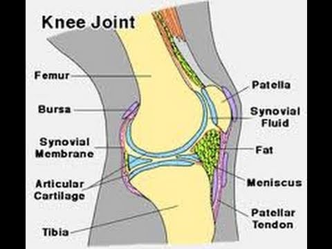 The study is co-authored by A. Nakamura, D. Murata, R. Fujimoto, S. Tamaki, S. Nagata, M. Ikeya, J. Toguchida, and K. Nakayama.
Subscribe to the 3D Printing Industry newsletter for the latest news in additive manufacturing. You can also stay connected by following us on Twitter and liking us on Facebook.
Looking for a career in additive manufacturing? Visit 3D Printing Jobs for a selection of roles in the industry.
Subscribe to our YouTube channel for the latest 3D printing video shorts, reviews and webinar replays.
Featured image shows schematic representation of the 3D printing process. Image via Biofabrication.
Tags A. Nakamura Central Queensland University Chalmers University of Technology Cyfuse Biomedical K.K. D. Murata FC Barcelona J. Toguchida K. Nakayama Kenzan Kyoto University M. Ikeya Pennsylvania State University R. Fujimoto Regenova S. Nagata S. Tamaki Saga University Scar Free Foundation swansea university TRIANKLE University of Alberta
Hayley Everett
Hayley is a Technology Journalist for 3DPI and has a background in B2B publications spanning manufacturing, tools and cycling. Writing news and features, she holds a keen interest in emerging technologies which are impacting the world we live in.
Human cartilage has been successfully 3D printed
3D printers have been causing revolutions in many different fields, with materials as different as food, mud, plastic, and plants. The game-changer is that you can create very precise, complex shapes that weren’t able to be created before. Another use of 3D printing is a potentially life-saving one. 3D bioprinters are being developed that can print out tissues and organs. Some that are being tested now are skin cells, bone, heart tissue, and now cartilage. A team of researchers at Sahlgrenska Academy has created cartilage tissue by printing stem cells with a 3D-bioprinter.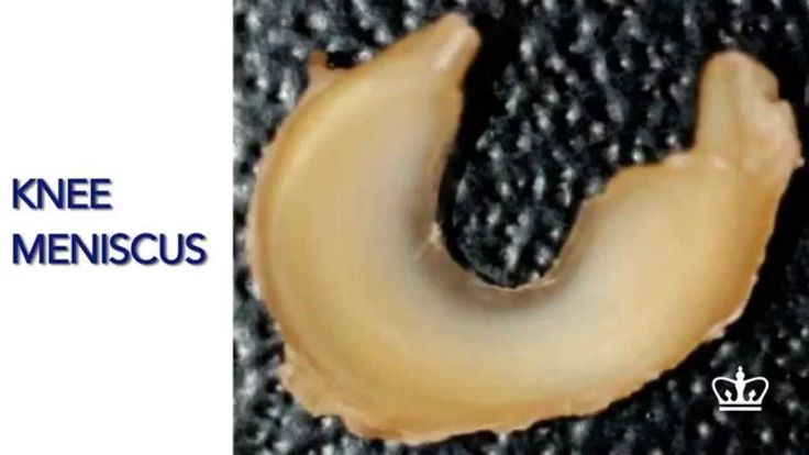 It appears to be just like human cartilage and could be used to replace damaged cartilage.
The lead researcher, Stina Simonsson, holding some 3D-printer cartilage. Image credits: Elin Lindström Claessen.“In nature, the differentiation of stem cells into cartilage is a simple process, but it’s much more complicated to accomplish in a test tube. We’re the first to succeed with it, and we did so without any animal testing whatsoever,” says Stina Simonsson, Associate Professor of Cell Biology, who led the research.
The researchers took cartilage cells from patients who had recently had knee surgery and their cells were manipulated to become “pluriplotent”, so they can develop into many different types of cells. Next, they created a scaffold to print the cells on. The stem cells were coated with nanocellulose to survive the printing process. Once printed, the stem cells multiplied and were given growth factors so they differentiated into cartilage tissues. The cells formed cartilage cells on the printed structure. After a few weeks, the cells lost their ability to change into other cells. This change is good because pluripotency increases the risk of tumour formation.
Cartilage can be 3D-printed. Image credits: United States NIH National Institute of Arthritis and Musculoskeletal and Skin Diseases.“We investigated various methods and combined different growth factors. Each individual stem cell is encased in nanocellulose, which allows it to survive the process of being printed into a 3D structure. We also harvested mediums from other cells that contain the signals that stem cells use to communicate with each other so called conditioned medium. In layman’s terms, our theory is that we managed to trick the cells into thinking that they aren’t alone,” says Stina Simonsson.
The 3D bio-printed structure is very similar to human cartilage. Experienced surgeons did not see a difference between natural and bio-printed cartilage.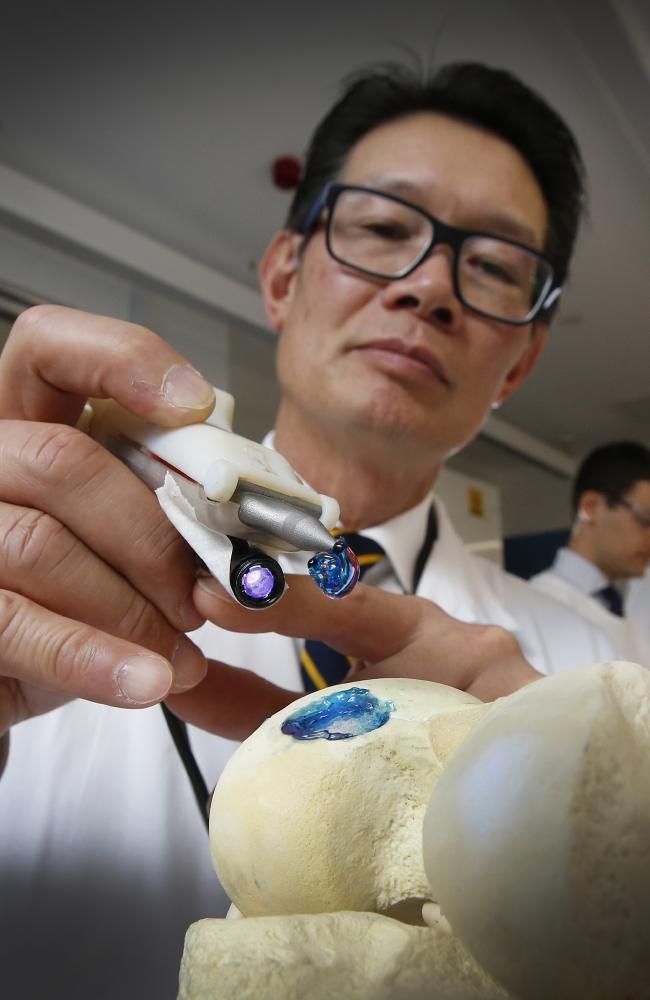 The cells appear well-formed under the microscope and similar to the patients’ own cartilage.
When this method is perfected, cartilage could be 3D printed from a patient’s own stem cells to repair damaged cartilage or heal osteoarthiritis (cartilage decay in the joints). The method can create a lot of cartilage, making it very useful for cartilage replacement. Right now, it isn’t known how compatible it is in the human body. The structural material needs to be able to break down and be absorbed safely by the body so only cartilage is left. Further development and testing need to be conducted. Bioprinting has a lot of potential; in the near future, tissues and organ could be printed on demand.
Journal reference: Nguyen, D. et al. 2017. Cartilage Tissue Engineering by the 3D Bioprinting of iPS Cells in a Nanocellulose/Alginate Bioink. Scientific Reports.
| 3DNews Technologies and IT market. News printers, print servers, scanners, copiers... The new material allows you to print knee... The most interesting in the reviews 05/02/2017 [14:13], Konstantin Khodakovsky Scientists from the American Duke University have created a material that mimics human cartilage and can eventually be used by surgeons for 3D printing of implants to replace damaged parts of the knee joint, individually shaped to the anatomy of each patient. nine0007 Human knees have a pair of menisci, crescent-shaped cartilages that act as shock absorbers. But over years of stress, these important parts of the joint can wear out, or be severely damaged by one wrong move while playing football or tennis. The result is pain and an increased risk of developing arthritis. Scientists claim their hydrogel-based material is the first to match the strength and elasticity of human cartilage while remaining stable inside the body and allowing it to be used in the 3D printing process. Duke University Associate Professor of Chemistry and Published Author Benjamin Wiley notes: “We have now made it possible for everyone to print implants that are very similar in medical properties to human cartilage in a fairly simple and relatively inexpensive way.” . It is worth noting that this is indeed an important achievement. The fact is that torn or damaged menisci are very capricious to the process of self-healing. Surgeons often have to partially or completely remove them, and existing implants either do not match the strength and elasticity of the original, or have poor biocompatibility and prevent the healing of the damaged area. nine0007 Recently, materials called hydrogels, which are very similar in molecular structure to cartilage and are biologically compatible, are increasingly being used as replacements for cartilage. Source: If you notice an error, select it with the mouse and press CTRL+ENTER. Related materials Permanent URL: https://3dnews.ru/951559 Headings: News Hardware, printers, print servers, scanners, copiers, MFPs, cutting edge science, Tags: 3d printer, 3d printing, surgery, medicine, science ← В past To the future → |
 To prove their point, the researchers used a $300 3D printer to print custom menisci for a plastic model of the knee. nine0007
 Doctor 3D prints knee cartilage
Archive
Follow author
Subscribe
Don't
Dr.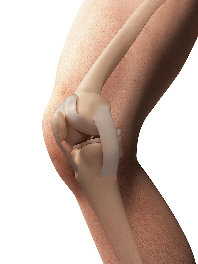 Darryl D'Lima and colleagues at La Scripps Clinic, California a new 3D bioprinting technology that will allow the implantation of living cartilage tissue into the patient's body, thereby providing better treatment for knee injuries. nine0007
Colleagues consider Dr. D'Lim a very resourceful person. He is a doctor and scientist who uses new technologies from various fields of science to help his patients.
Recently, Dr. D'Lima has been working on a technology for implanting 3D-printed cartilage tissue into the patient's body to restore the structure of damaged knees. Cartilage is the tissue that connects the knee joints. It is known that it heals slowly and painfully.
Now doctors advise patients to endure the pain if there is no need for an artificial joint. This procedure also causes severe pain and rarely solves the problem.
Hoping to find an alternative to conventional joint implants, Dr. D'Lima and his team decided to make a 3D bioprinter out of an old HP inkjet printer and print living cartilage tissue on it.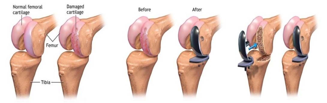 The 3D printer sprays a mixture of cartilage progenitor cells and a liquid that hardens under ultraviolet light. Bone cells are also printed on it, which are placed where the cartilage connects to the bone. The volume of a drop of liquid with living cells is 1 picoliter, which is quite enough to correct even microscopic disorders of the patient's bone or cartilage. nine0007
Dr. D'Lima compares the process of implanting cartilage into the knee with filling in potholes: “You automatically fill in the depressions during printing. You get cartilage that is perfectly shaped to the existing tissue, which we have never been able to do with conventional implants,” said D’Lima. Printing on a pre-existing joint provides a better fit for new and old cartilage than grafting lab-grown cartilage that has to be trimmed to fit better. Another advantage of cartilage printing is that there is no longer a need to postpone the operation for later - it can be done right away. nine0007
People born during the "baby boom" age, and the problem of excess weight is becoming more and more aggravated. This means that in the future, knee injuries will become an even more serious cause for concern. According to a GlobalData report, the knee implant market will grow by 6.8% annually from 2010 to 2017.
Dr. D'Lim and his team's research has been recognized by several institutes. He received a $3.1 million grant from the California Institute for Regenerative Medicine Stem Cell Research Agency to research the use of natural and artificial embryonic stem cells in cartilage growth. nine0007
D'Lima believes that it will take at least a year to create a full-fledged 3D bioprinter. He is currently conducting animal experiments to prove the promise of his idea, which is sponsored by the George Schaeffer Family Foundation.
Similar 3D bioprinting inventions are popping up around the world. Just last month, a prototype 3D printed BioPen was handed over to researchers at St Vincent's Hospital in Melbourne, and Organovo created the world's first 3D printed liver tissue on a printer in its labs.