3D printer human tissue
3D printing cells to produce human tissue • tectales • tagging medical technology
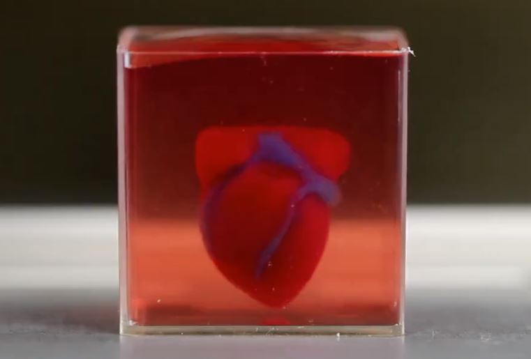
University of Utah biomedical engineering assistant professor Robby Bowles and his team have developed a method to 3D print cells to produce human tissue such as ligaments and tendons to greatly improve a patient's recovery.
Source: Dan Hixson/University of Utah College of Engineering
17.10.2018 •
- #3D printing
- #bioprinting
- #hydrogel
Engineers have developed a method to 3D print cells to produce human tissue such as ligaments and tendons, a process that will greatly improve a patient's recovery.
With today’s technology, we can 3D print sculptures, mechanical parts, prosthetics, even guns and food. But a team of University of Utah biomedical engineers have developed a method to 3D print cells to produce human tissue such as ligaments and tendons, a process that will greatly improve a patient’s recovery. A person with a badly damaged ligament, tendon, or ruptured disc could simply have new replacement tissue printed and ultimately implanted in the damaged area. “It will allow patients to receive replacement tissues without additional surgeries and without having to harvest tissue from other sites, which has its own source of problems,” says University of Utah biomedical engineering assistant professor Robby Bowles, who co-authored the paper along with former U biomedical engineering master’s student, David Ede.
The 3D printing method, which took two years to research, involves taking stem cells from the patient’s own body fat and printing them on a layer of hydrogel to form a tendon or ligament which would later grow in vitro in a culture before being implanted. But it’s an extremely complicated process because that kind of connective tissue is made up of different cells in complex patterns. For example, cells that make up the tendon or ligament must then gradually shift to bone cells so the tissue can attach to the bone. “This is a technique in a very controlled manner to create a pattern and organizations of cells that you couldn’t create with previous technologies,” Bowles says of the printing process. “It allows us to very specifically put cells where we want them.”
This image of cells that were made fluorescent shows how they are printed in complex structures for the purpose of producing tissue such as tendons and ligaments.
Source: Robby Bowles/ University of Utah College of Engineering.
To do that, Bowles and his team worked with Salt Lake City-based company, Carterra, Inc., which develops microfluidic devices for medicine. Researchers used a 3D printer from Carterra typically used to print antibodies for cancer screening applications. But Bowles’ team developed a special printhead for the printer that can lay down human cells in the controlled manner they require. To prove the concept, the team printed out genetically-modified cells that glow a fluorescent color so they can visualize the final product.
Currently, replacement tissue for patients can be harvested from another part of the patient’s body or sometimes from a cadaver, but they may be of poor quality.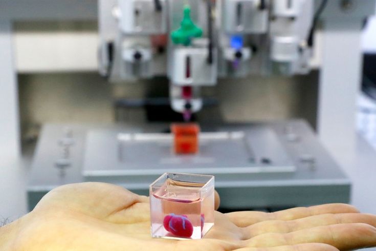 Spinal discs are complicated structures with bony interfaces that must be recreated to be successfully transplanted. This 3D-printing technique can solve those problems.
Bowles, who specializes in musculoskeletal research, said the technology currently is designed for creating ligaments, tendons and spinal discs, but “it literally could be used for any type of tissue engineering application,” he says. It also could be applied to the 3D printing of whole organs, an idea researchers have been studying for years. Bowles also says the technology in the printhead could be adapted for any kind of 3D printer.
- #3D printing
- #bioprinting
- #hydrogel
- #materials
- #microfluidics
- #surgery
- #tissue
Source: University of Utah
Rapid new bioprinting method unlocks potential of human tissue transplants
0Shares
Scientists from the University at Buffalo have developed a rapid new 3D bioprinting method that could represent a significant step towards fully-printed human organs.
Using a novel vat-SLA-based approach, the team have been able to reduce the time it takes to create cell-laden hydrogel structures, from over 6 hours to just 19 minutes. The expedited biofabrication method also enables the production of embedded blood vessel networks, potentially making it a significant step towards the lifesaving 3D printed organs needed by those on transplant waiting lists.
“Our method allows for the rapid printing of centimeter-sized hydrogel models,” explained the study’s lead co-author, Chi Zhou. “It significantly reduces part deformation and cellular injuries caused by the prolonged exposure to the environmental stresses you commonly see in conventional 3D printing.”
“The technology we’ve developed is 10-50 times faster than the industry standard, and it works with large sample sizes that have been very difficult to achieve previously.”
Taking bioprinting up a gear
Although bioprinted cell‐laden structures hold significant potential when it comes to human tissue and organ transplants, the technology is still at a nascent stage. One of the main hurdles facing the wider adoption of these processes is print speed, as the deposition rates of hydrogels have so far been limited to avoid damaging their incumbent cells.
Nozzle-based techniques have other drawbacks too, as they can cause prolonged cellular exposure to shear stress as well as low oxygen levels and temperatures, damaging them in the process. What’s more, the hydrogel scaffolds produced using conventional methods often exhibit low mechanical strength, making it difficult to incorporate soft overhanging structures like vascular channels.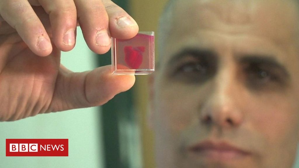
While using sacrificial supports enables scientists to partially overcome this deficiency, the simplicity of the extrusion method behind this approach continues to limit its capacity. By contrast, recently-developed Continuous Liquid Interface Production (CLIP) technologies, have the potential to drastically increase the speed of bioprinting processes.
By continuously building layers above a ‘dead zone,’ CLIP methods allow materials to constantly be replenished, increasing production capacity, but at the cost of only being able to create thin-walled parts. Building on this approach, the Buffalo team have now developed a ‘FLOAT’ method, in which hydrogels can be deposited at a higher velocity, enabling the production of larger vascularized tissues.
The scientists were able to 3D print a hand-shaped structure (pictured) in less than 20 minutes. Photo via the Advanced Healthcare Materials journal.The ‘FLOAT’ bioprinting approach
During the researchers’ optimized FLOAT method, objects are essentially cured through a glass plate inside a vat of hydrogel at low suction forces, yielding thick parts with high elasticity. To prove the biocompatibility of their approach, the team initially fabricated a set of specimens from the cell-compatible PEGNB polymer.
Interestingly, while test parts exhibited sufficient rigidity, they also shrunk by up to 51%, causing the researchers to switch to a PEGDA material for larger models. In more ambitious test runs, the Buffalo team then 3D printed several 2.6 × 1.7 × 5.6 cm hand-shaped hydrogel structures, with ‘fingers’ that bent under compression.
Producing the same models using a normal SLA 3D printer took the team around 6.5 hours, significantly longer than the 19 minutes of their FLOAT-based machine. The scientists’ hydrogel-based hands also featured vascular channels, meaning that they could eventually be seeded with endothelial cells to create functional, transplantable limbs.
Ultimately, the scientists were able to seed patches of cells into microchannels ex-vivo, but they also found that integrating these into higher-strength structures yielded low cell viability. In future, the team believe that switching to nanomaterial-doped polymers could provide the answer to balancing rigidity and compatibility, and enable the rapid production of hydrogel-based vascularized structures.
Inching bioprinting towards reality
While the 3D bioprinting remains at a largely experimental stage, there are signs that the technology is slowly progressing towards more end-use applications.
3D printer OEM 3D Systems announced a major breakthrough in its Print to Perfusion bioprinting platform earlier this year. The system is now capable of creating fully-sized vascularized lung scaffolds, and the company has indicated that the technology will soon play a key role within its healthcare business.
Biotechnology firm United Therapeutics and Israeli company CollPlant have also made significant advances in their bid to mass-manufacture 3D printed kidneys. The companies have turned a former tobacco factory into a modern 3D bioprinting production line, which could be capable of churning out additive organs.
Elsewhere, efforts to create functional human organs have been limited to miniaturized models, such as the tiny 3D bioprinted hearts created by scientists at the University of Texas at El Paso. The vascularized structures were sent to the International Space Station (ISS) to test how microgravity affects the human heart.
The researchers’ findings are detailed in their paper titled “Fast Stereolithography Printing of Large‐Scale Biocompatible Hydrogel Models. ” The study was co-authored by Nanditha Anandakrishnan, Hang Ye, Zipeng Guo, Zhaowei Chen, Kyle I. Mentkowski, Jennifer K. Lang, Nika Rajabian, Stelios T. Andreadis, Zhen Ma, Joseph A. Spernyak, Jonathan F. Lovell, Depeng Wang, Jun Xia, Chi Zhou and Ruogang Zhao.
To stay up to date with the latest 3D printing news, don’t forget to subscribe to the 3D Printing Industry newsletter or follow us on Twitter or liking our page on Facebook.
Are you looking for a job in the additive manufacturing industry? Visit 3D Printing Jobs for a selection of roles in the industry.![]()
Featured image shows a University at Buffalo 3D bioprinted hand. Photo via the Advanced Healthcare Materials journal.
Tags 3D Systems Chi Zhou CollPlant International Space Station United Therapeutics University at Buffalo University of Texas at El Paso
Paul Hanaphy
Paul is a history and journalism graduate with a passion for finding the latest scoop in technology news.
Scientists develop a 3D printer model for printing bone implants
Media about this project already told in the spring. New development will fill the voids in human bones with a material based on the mineral hydroxyapatite, which will temporarily replace living tissue, will give impetus to the development own cells, and then dissolve, leaving behind healed cavity.
Natalya Bulina
What happens to the project after a year? Natalya Bulina, senior researcher of ICTTM SB RAS, spoke about the progress of work.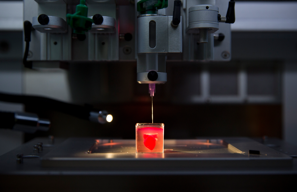
Despite the difficulties associated with the coronavirus, scientists have achieved impressive results. Already conducted in vitro and in vivo biological testing of apatites with different formulations. The studies were carried out in parallel: so far at the NIOCH SB RAS powder was implanted into the defects of the skull of rats, the Vector studied the action of the same substances on human bone tissue cells. During experiments, scientists determined the composition of apatite, which is the most effective both “in vitro” and on living organisms — stimulates the formation of new bone tissue and accelerates implantation implantable material. At the same time, native bone tissue cells people do not die, but actively multiply.
It is planned that the developed 3D printer will print on selective laser melting technology, so scientists tested the effect of laser radiation on the resulting composition apatite. It turned out that when the powder is melted, the substance does not is destroyed, and while the implant will be printed, it will not lose their healing properties.
The successful completion of this stage of work was the publication research results in Ceramics magazine international.
IA&E SB RAS is another executor of the project together with ICTTM SO RAS — developed software and control module the main nodes of the created 3D printer layout. In that year, scientists will develop a layer-by-layer printing unit - sequential deposition of layers of hydroxyapatite on top of each other.
In medicine, titanium implants printed on 3D printer. They are used in maxillofacial surgery, traumatology, orthopedics, oncology. These foreign of the human body, the products are fixed in bone defects with screws and remain in the body forever. In IHTTM pursue a different goal - the material from which it will be printed the product will eventually dissolve in the human body and will turn into its own bone.
Hydroxyapatite, synthesized in powder form
— Hydroxyapatite is a source of phosphorus and calcium, from which then our bone tissue is formed. And the additives introduced into the structure of hydroxyapatite in low concentration, necessary to accelerate the process of degeneration of the implant into native bone tissue,” explains Natalia Bulina.
This material is suitable for the restoration of small bone defects that do not carry a strong load - this is mainly Maxillofacial Surgery. In addition, they can fill cavities and cracks in the bones after serious illnesses and injuries. printed products will be individual: they must be designed from tomography data of a particular person.
Natalya believes that for the effective implementation of the synthesized hydroxyapatite between a synthesis specialist and a doctor should be an intermediate link that will modernize received material for a specific medical task and will be able to work individually with each specific case:
— To date, we have developed a technology for obtaining so-called raw material and we can give it, and make it ready medical products for a specific medical problem only with the direct participation of physicians.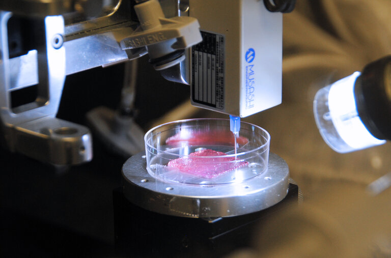 In addition, this medical products must be tested on living organisms, And these are long-term tests, from a month to a year. Therefore, the implementation of the hydroxyapatite we synthesize is a slow process.
Scientists hope that the 3D printer model being developed will in demand on the market, and ongoing research will expand opportunities for the use of synthetic hydroxyapatite in medicine.
Information and photos are provided by the press service of IHTTM SO RAS
Scientists printed human skin on a 3D printer (video)
The Chinese have learned how to install a SIM card tray in the iPhone 14 without a SIM card
Chinese enthusiasts disassembled the iPhone 14, brought from the US, and found that instead of the SIM card tray, the new SIM-free iPhone 14 for the US has a plastic plug. It was decided to conduct an experiment on the "implantation" of the SIM card slot.
Read moreBright photo portraits and stylish design: vivo introduces the V25 Series in Russia (3 photos)
Vivo announces the launch of the new V25 Series on the Russian market, introducing the new V25 Pro with a 120 Hz curved 3D screen, V25 and V25e with a 64 MP ultra-sensitive camera and hybrid stabilization.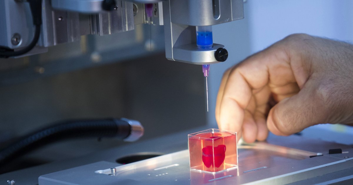 Stylish smartphones with FlashCharge fast charging, 8 GB virtual memory expansion and photochromic glass body are available in various colors...
Benefits of antivirus software
If you still have not wondered why install antivirus software, what is the use of it, then you should do it as soon as possible. Because only such software can ensure the full security of your computer or mobile device, prevent malware from penetrating...
Read moreiPhone and Android learned how to hack through the WhatsApp messenger
WhatsApp developers have admitted to finding a serious vulnerability in the application code. It turned out that in the latest versions of WhatsApp, hackers could hack the messenger by receiving a video call from the user, while watching the video also threatens to be hacked, because hackers began to “sew” viruses into the video.
Read moreSeries Infinix NOTE 12 will be replenished with a new model (3 photos)
Infinix has announced the imminent release of the new NOTE 12 2023 (G99). The novelty will join the range of high-performance Infinix devices and is aimed at users who value multitasking, as well as mobile gaming.
Vkontakte Odnoklassniki
A team from the Carlos III University of Madrid, the Center for Research in Energy, Environment and Technology (CIEMAT), the Gregorio Marañon University Hospital of Madrid and the BioDan Group have collaborated to develop a 3D bioprinter that creates an artificial skin that mimics human structure. The complexity of its development was to establish a methodology in which correctly selected proportions, mixing and sequential application of biological materials made it possible to obtain a functional tissue.
3D printed skin consists of epidermis and dermis. The samples are biologically active, nourish, renew, produce collagen, which ensures the elasticity of the tissue. The resulting skin is quite suitable for transplantation in patients with severe burns.