Nih 3d print exchange
The NIH 3D Print Exchange: A Public Resource for Bioscientific and Biomedical 3D Prints
- Journal List
- Liebert Funded Articles
- PMC4981148
3d Printing and Additive Manufacturing
3D Print Addit Manuf. 2014 Sep 1; 1(3): 137–140.
doi: 10.1089/3dp.2014.1503
PMCID: PMC4981148
NIHMSID: NIHMS810734
PMID: 28367477
,1,*,1,*,1,1,2,3,4,5,3,2,4 and 1
Author information Copyright and License information Disclaimer
The National Institutes of Health (NIH) has launched the NIH 3D Print Exchange, an online portal for discovering and creating bioscientifically relevant 3D models suitable for 3D printing, to provide both researchers and educators with a trusted source to discover accurate and informative models. There are a number of online resources for 3D prints, but there is a paucity of scientific models, and the expertise required to generate and validate such models remains a barrier. The NIH 3D Print Exchange fills this gap by providing novel, web-based tools that empower users with the ability to create ready-to-print 3D files from molecular structure data, microscopy image stacks, and computed tomography scan data. The NIH 3D Print Exchange facilitates open data sharing in a community-driven environment, and also includes various interactive features, as well as information and tutorials on 3D modeling software. As the first government-sponsored website dedicated to 3D printing, the NIH 3D Print Exchange is an important step forward to bringing 3D printing to the mainstream for scientific research and education.
The NIH 3D Print Exchange (“the Exchange”) is an online portal from the National Institutes of Health (NIH) for discovering, creating, and sharing bioscientific 3D models that are ready to download and print in 3D (). The Exchange provides a publicly accessible venue for a community of users to share their innovative 3D models with the global community, and to learn and discuss new ways to design and utilize bioscientific 3D prints.
Open in a separate window
The NIH 3D Print Exchange is a virtual collection of bioscientific 3D models and tutorials for 3D printing, sponsored by the National Institutes of Health. Scan the QR code to visit the web site
The Exchange began a public “beta” at the USA Science and Engineering Festival, held in Washington, DC, on April 26–28, 2014. The official release was announced on June 18 in conjunction with the White House Maker Faire, where the Exchange was a featured exhibit. Within one month, over 11,000 unique visitors had viewed the site.
Tangible models built by 3D printers are highly valuable in biomedical research and communication as well as education,1–3 and 3D visualization and printing has facilitated scientific insights and changed the direction of biomedical research (). In medicine, 3D prints are being used in surgical planning and in creating custom implants and prosthetics.4 Bioprinting and tissue fabrication methods are rapidly advancing, and the potential is vast.5–7 At the NIH, 3D printing has saved hundreds of research animals and precious samples in some of the most advanced labs in the country; we estimate that tens of thousands of dollars in research funding have been saved by streamlining laboratory processes and avoiding downtime, as well as saving researchers' time and speeding the path to discovery.
Open in a separate window
Physical representations of complex structures can provide valuable insights into otherwise unseen features. A 3D print of hemagglutinin (3DPX-000027), a receptor on the influenza virus, helped change the direction of research toward a universal flu vaccine (print by Darrell Hurt, photo by Jeremy Swan).
Despite the fast-growing applications for 3D printing, it is not widely used in the biosciences at present. With the exception of well-trained modeling specialists and 3D printing enthusiasts, most researchers and lay people have little to no experience using the software required to create digital models. 3D printers are affordable and readily accessible, and hundreds of thousands of printable models can be found online. However, in an online sea of gadgets, toys, and accessories numbering into the thousands, there were few models relevant to scientific research, medical practice, or STEM (science, technology, engineering, and mathematics) education.
The Exchange thus fills an important, latent need: to provide scientifically accurate, high-quality 3D models in a ready-to-print format, bridging the knowledge gap between a novice user and the 3D modeling specialist. The site's novel, open source tools generate 3D printable models of molecular, microscopy, and medical imaging data through automated pipelines (). What used to take hours for a skilled user is now possible in only minutes by even the most naïve user. By democratizing access to bioscientific 3D models on demand, the Exchange enables users of all levels to focus their time less on tedious tasks and more on research and discovery, while at the same time vastly increasing the number of 3D printable models available to the bioscientific research community.
Open in a separate window
Novel web-based tools generate STL and VRML files, each with a corresponding PNG image and X3D version. Molecular structures are output in seven variants; examples from entry 3DPX-000479 show (A) ribbon secondary structure and (B) hydrophobic surface of a protein in complex with DNA (A, green; B, pink).10 Isosurfaces derived from microscopy data and image stacks are rendered in monochrome and with radial coloring, (C) the latter shown for Human Papilloma Virus (EMDB-5839),11 3DPX-000406.
In addition to 3D models and free tools, the Exchange brings added value by providing video tutorials for 3D modeling software, with a focus on converting medical and scientific data.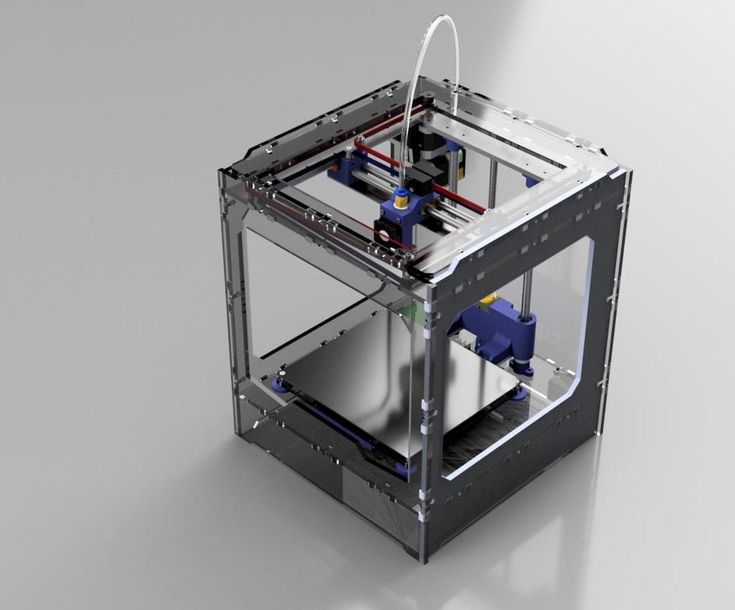 These are intended to empower users with the skills to customize their own models, beyond the basic offerings in the automated pipelines. The site also hosts a discussion forum to cultivate a community of users and to facilitate idea sharing and collaboration among them.
The Exchange provides both researchers and educators with a trusted source to discover accurate and informative models. Because models can be generated from data derived directly from scientific databases (the Protein Data Bank8 and the Electron Microscopy Data Bank9), users can expect a higher level of confidence in scientific accuracy. All models are given a unique ID number (e.g., 3DPX-123456), so they can be cited in scientific publications and referenced across the web and in social media.
Next steps for the Exchange are to optimize the automated tools that convert scientific and medical data to ready-to-print stereolithography (STL) and virtual reality modeling language (VRML) file formats, as well as extensible 3D graphics (X3D) files that are utilized in the in-browser X3DOM12 viewer (). Improvements to these pipelines will focus on increasing model quality, improving data compression to facilitate faster downloads, and accommodating more data types, such as magnetic resonance imaging (MRI) or positron emission tomography (PET) scans for medical images. Because open source development and the principles of open data are part of the overarching mission of the Exchange, the code to these tools will be placed in the public domain, and web services will be implemented.
Open in a separate window
A screenshot of the X3DOM viewer. An X3D was autogenerated from an STL (3DPX-000404) uploaded by Dr. Bruno B. Gobbato (username bgobbato), shoulder and elbow specialist, Instituto de Ortopedia e Traumatologia de Jaraguá do Sul, SC, Brazil. Model based on computed tomography scan data of the lower cranium and cervical vertebrae.
The Exchange is built on Drupal, an open source content management system facilitating free and easy transfer of the site framework to other federal agencies. In this way, the Exchange supports the government movement to make scientific collections, including digital 3D data, freely available to the public.11 The system could be adopted to enable other exchanges, for example, 3Dprint.agency.gov or 3Dprint.university.edu.
The community-driven aspect of the Exchange is critical to its success, and we invite users to share models related to medicine and biosciences. To that end, we are continuing to enhance the user interface and user experience, primarily through feedback from the ever-growing user community. We will also be implementing specially curated “collections” and encouraging the use of 3D prints in science education by soliciting users to upload their own supplemental materials to be used in the classroom alongside 3D-printed models.
3D printing technology is advancing rapidly, with the expectation that within the next decade, 3D-printed human tissues and organs will regularly be used in medical treatment. 13 The Exchange is thus a well-positioned resource for supporting this significant medical development, and puts the NIH and the U.S. Department of Health and Human Services ahead of this emerging technology, which aligns with their interests to promote research leading to new and improved treatments for patient care. Furthermore, the Exchange supports government initiatives in the Maker Movement and STEM education.14 Ultimately, we hope that the NIH 3D Print Exchange will help to bolster the use of 3D printing in medical and bioscientific research, education, and communication.
The Exchange is owned and operated by the National Institute of Allergy and Infectious Diseases under guidance from Mr. Mike Tartakovsky, chief information officer. The Exchange is funded in part by HHS Ignite and HHS Ventures, initiatives sponsored by the HHS IDEA Lab of the Office of Technology, U.S. Department of Health and Human Services. The Exchange gratefully acknowledges the time and feedback from members of our Advisory Team.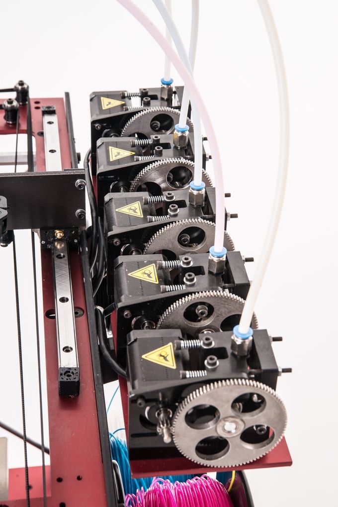 Special recognition goes to Tom Ferrin, PhD, and the Chimera Team at the University of California San Francisco for help in optimizing 3D modeling workflows, and Nicholas Polys, PhD, Virginia Tech, for X3D and WebGL development support. For valuable feedback and special contributions, we also thank the NIH Library Office of Research Services, Eric Jones and Sam Michael of the National Center for Advancing Translational Sciences, the White House Office of Science and Technology Policy, and Jordan Miller, PhD, Rice University.
The authors declare no competing financial interests exist. Reference to any specific persons, commercial products, process, or service by trade name, trademark, manufacturer, or otherwise does not necessarily constitute or imply endorsement, recommendation, or favoring by the U.S. Government. The views and opinions of authors expressed herein do not necessarily state or reflect those of the U.S. Government, and shall not be used for advertising or product endorsement purposes.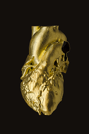
1. Kolitsky M. 3d printed tactile learning objects: proof of concept. J Blindness Innovation Res 2014;4(1) [Google Scholar]
2. McMenamin P, Quayle M, McHenry C, et al.. The production of anatomical teaching resources using three-dimensional (3D) printing technology. Anat Sci Ed 2014; doi: 10.1002/ase.1475 [PubMed] [CrossRef] [Google Scholar]
3. Costello JP, Olivieri LJ, Krieger A, et al.. Utilizing three-dimensional printing technology to assess the feasibility of high-fidelity synthetic ventricular septal defect models for simulation in medical education. World J Pediatr Congenit Heart Surg 2014;5:421–426 [PubMed] [Google Scholar]
4. Kung TA, Bueno RA, Alkhalefah GK, et al.. Innovations in prosthetic interfaces for the upper extremity. Plast Reconstr Surg 2013;132:1515–1523 [PubMed] [Google Scholar]
5. Cui X, Gao G, Yonezawa T, et al.. Human cartilage tissue fabrication using three-dimensional inkjet printing technology. J Vis Exp 2014; doi: 10.3791/51294 [PMC free article] [PubMed] [CrossRef] [Google Scholar]
6.![]() Miller JS. The billion cell construct: will three-dimensional printing get us there? PLoS Biol 2014;12:e1001882. [PMC free article] [PubMed] [Google Scholar]
Miller JS. The billion cell construct: will three-dimensional printing get us there? PLoS Biol 2014;12:e1001882. [PMC free article] [PubMed] [Google Scholar]
7. Murphy SV. and Atala A. 3D bioprinting of tissues and organs. Nat Biotech 2014;32:773–785 [PubMed] [Google Scholar]
8. Berman HM, Westbrook J, Feng Z, et al.. The Protein Data Bank. Nucleic Acids Res 2000;28:235–242 [PMC free article] [PubMed] [Google Scholar]
9. Lawson CL, Baker ML, Best C, et al.. EMDataBank.org: unified data resource for CryoEM. Nucleic Acids Res 2011;39(suppl 1):D456–D464 [PMC free article] [PubMed] [Google Scholar]
10. Wang Z, Wu Y, Li L, et al.. Intermolecular recognition revealed by the complex structure of human CLOCK-BMAL1 basic helix-loop-helix domains with E-box DNA. Cell Res 2013;23:213–224 [PMC free article] [PubMed] [Google Scholar]
11. Zhao Q, Potter CS, Carragher B, et al.. Characterization of virus-like particles in GARDASIL(R) by cryo transmission electron microscopy. Hum Vaccin Immunother 2013;10(3) [PMC free article] [PubMed] [Google Scholar]
12. The White House Office of the Press Secretary. FACT SHEET: President Obama to Host First-Ever White House Maker Faire. 2014. www.whitehouse.gov/the-press-office/2014/06/18/fact-sheet-president-obama-host-first-ever-white-house-maker-faire (last accessed August10, 2014)
13. Memorandum for the Heads of Executive Departments and Agencies: Improving the Management of and Access to Scientific Collections. 2014. www.x3dom.org/
14. Behr J, Eschler P, Jung Y, et al.. X3DOM: a DOM-based HTML5/X3D integration model. In: Proceedings of Web3D 2009: The 14th International Conference on Web3D Technology ACM, New York, NY, 2009; pp. 127–135 [Google Scholar]
NIH Has Open Source 3D Printed Medical Models
0Shares
You know how you’re always saying that everything should be free? Especially science stuff? Maybe you’re not saying that, unless you’re broke, but it’s definitely something that I shout at ads on the TV. Finally, my (possibly your) wants are starting to be met, at least in the realm of science. The USA’s National Institute of Health (NIH) has created an online platform for the exchange of 3D printable biomedical files.
The National Institute of Allergy and Infectious Diseases, in partnership with the Eunice Kennedy Shriver National Institute for Child Health and Human Development and the National Library of Medicine have created the NIH 3D Print Exchange. And, so far, the public beta site is awesome! It’s filled with free, 3D printable models of all of your favorite viruses, proteins and simple lab equipment. Want a 3D model of e.coli? Or how about an Agarose gel comb
for Electrophoresis? See the myriad of models already up on the site in the promo reel below:
If you feel as though the 3D Print Exchange is missing the molecular structure of your recently discovered macromolecule, you can design and upload your own printable files. Of course, we can’t expect every doctor/scientist/educator to know how to create CAD models of such things, so the NIH is trying to make it as simple as possible. The site will have prewritten scripts that can turn 3D medical data, such as free models from the Electron Microscopy Database, into 3D printable files using the open-source UCSF Chimera software package.
“Hey!” someone might say, “I’m not a doctor or a scientist. What’s the point of all of this free, 3D printable biomedical stuff?” These biomedical models, at the moment, may be great for educating students about a variety of chemical or anatomical structures. Moreover, doctors researching diseases and potential cures can 3D print models to communicate to other researchers about the makeup of a given virus or medicine so that they can understand it at a tangible level. As users expand the catalogue of medical models through 3D scanning, doctors dealing with a medical problem, such as a unique heart deformity, can review the online database for similar cases. And the lab equipment uploaded to the Exchange can be customized to fit the needs of specific labs so that, rather than order expensive components, such as an LED filter adapter from a supplier, they can obtain it more quickly and cheaply via 3D printing. The platform also paves the way for future technological developments that may take place. In a world where medicine, medical devices and prosthetics are all 3D printable, users may be able to go to a site like 3D Print Exchange and share files. NIH gets into the uses of 3D printing in medicine and their new site below:
All of the benefits of the NIH site are magnified for those in developing countries. 3D printable models for education may be more affordable than those purchased from manufacturers and access to knowledge produced by leading research institutions may become increasingly more accessible. When the site does begin to introduce 3D printable medical devices and the like, users in remote areas will be able to 3D print objects like prostheses for their local populations.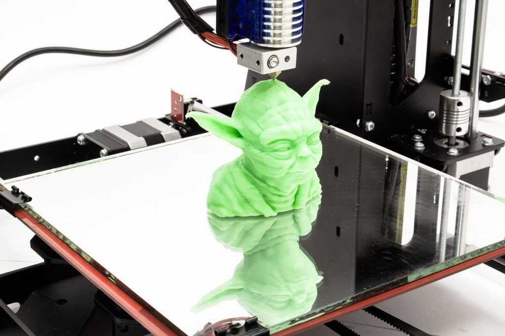
This is very exciting stuff! I can’t wait for Pfizer to get the hint and upload their chemical formulas to the NIH 3D Print Exchange. [Ed: It may take a while, Pfizer is currently too busy aiming for pharma world domination in its bid to acquire Astra Zeneca!]
Hat tip to Pjotr du Mât on Facebook.
Source: NIH 3D Print Exchange
Michael Molitch-Hou
Michael Molitch-Hou previously served as Editor-in-Chief of 3D Printing Industry, he is now the Editor of Engineering . com's 3D printing section. He has covered additive manufacturing technology day in and day out since 2012 and has hundreds of article to his credit. He is the founder of The Reality Institute.
Medical 3D printer overview
Use of 3D printing in medicine
Source: docwirenews.com
3D printing has been used in medicine since the early 2000s, when this technology was first used to make dental implants. Since then, the use of 3D printing in medicine has expanded significantly, with doctors around the world describing ways to use 3D printing to produce ears, skeletal parts, airways, jawbones, eye parts, cell cultures, stem cells, blood vessels and vasculature, tissues and organs, new dosage forms and much more.
Source: zortrax.com
Using files with models for 3D printing provides an opportunity for the exchange of work among researchers. Instead of trying to reproduce the parameters described in scientific journals, doctors can use and modify ready-made 3D models. To this end, in 2014, the National Institutes of Health established the 3dprint.nih.gov exchange to facilitate the exchange of open source 3D models for medical and anatomical devices, custom equipment, and mock-ups of proteins, viruses, and bacteria.
Source: 3dprint.com
Modern medical use of 3D printing can be divided into several broad categories: tissue and organ fabrication, prostheses, implants and anatomical models, instrument printing, and pharmaceutical research.
Top five uses for 3D printing in medicine
Operational preparation and student education
Source: 3dprint.com
Taking into account individual differences and features of the anatomy of a particular human body, 3D printed models can be used to prepare surgical operations. Having a doctor have a tangible model of a particular patient's organ, made, for example, based on the results of CT (computed tomography) for study or to simulate an operation, significantly reduces the risk of medical errors.
Source: openbiomedical.org
The use of 3D models for training surgeons and students is preferable to training on cadavers, as it does not create problems in terms of availability and cost of objects. Cadavers often lack appropriate pathology, so they are more suitable for anatomy lessons than for presenting a patient with a disorder appropriate to the topic under study. With the help of 3D printing, it is possible to create a model of any organ with any known pathology.
Source: ncbi.nlm.nih.gov
two-dimensional images.
Bioprinting of tissues and organs
Source: hbr.org
Bioprinting is one of the many types of 3D printing used in the medical field. Instead of printing using plastic or metal, bioprinters use a syringe dispenser to apply bioink (layers of living cells or a structuring base for them) to create artificial living tissue. In addition to being used as an alternative to donor tissues, such tissue constructs or organoids can be used for medical research.
Source: press.ginkgo3d.com
Although 3D bioprinting systems can be laser, inkjet, or extrusion, inkjet bioprinting is the most common. Multiple printheads can be used to accommodate different types of cells (organ-specific, blood vessel cells, muscle tissue), which is a major challenge in the fabrication of heterocellular tissues and organs. 3D printing with biological materials can be used to regenerate tissues, and in the future, organs, directly on the patient.
Printing Surgical Instruments
Volt Grip Details, Source: bitegroup.nl
Today's surgeons are trying to perform operations with as little trauma to the patient as possible, so they very often require a personalized instrument. The use of 3D printing makes it possible to create such tools within hours.
Volt capture model visualization, Source: bitegroup.nl
Now the doctor can independently modify the finished model, giving it the necessary size and shape for convenience and efficiency.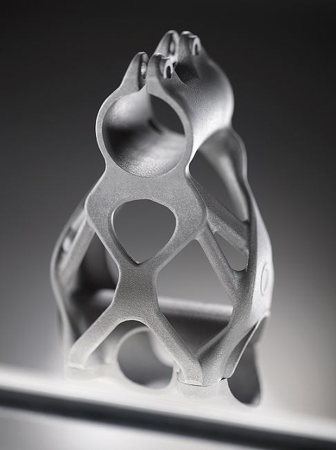 Dentists can now create, for example, individual guides right in front of the patient, eliminating the possibility of damage to healthy teeth during prosthetics.
About the clamp Volt, from the photos above, read further in the section “Examples of use”.
Here's how students at Duke University in Durham, North Carolina create tools using metal 3D printing.
"Printing" drugs
Source: mdpi.com
3D printing technologies are already being used in pharmaceutical research and personalized medicine, and their scope is constantly expanding. 3D printing enables precise dose control of drugs and the production of dosage forms with complex drug release profiles and prolonged action. Now pharmacists can analyze a patient's pharmacogenetic profile and other characteristics such as age, weight, or gender to determine the optimal dose and sequence of medications. If necessary, the dose may be adjusted, depending on the clinical response. With 3D printing, it is possible to produce personalized medicines in completely new formulations, such as tablets containing multiple active ingredients, either as a single mixture or as complex multi-layered tablets.
Prosthetics and Dentistry
Source: eos.info
3D printing has been successfully used in medicine for the manufacture of complex custom prostheses or surgical implants. Implants and prostheses of any possible geometry can be made by converting X-ray, MRI or CT images into a 3D printable model using special software.
The rapid production of custom implants and prostheses solves a pressing problem in orthopedics, where standard implants often do not fit the patient. This is also true in neurosurgery: skulls are individually shaped, so it is difficult to standardize a cranial implant. Previously, surgeons had to use various tools to modify and fit implants, sometimes right during the operation. The use of 3D printers makes this procedure unnecessary. Additive technologies are especially in demand when it is necessary to urgently manufacture implants.
A real revolution in dentistry came with the advent of 3D technology.
Source: hypowerfuel.com
First, complete and accurate 3D scanning of the oral cavity is now possible. Secondly, the use of 3D printing has made it possible to create prostheses that absolutely fit the anatomy of the patient, without the need for a long and unpleasant fit. The radical reduction in the share of manual labor in the manufacture of prostheses or veneers has reduced the required tolerances in production, expanded the list of materials used and increased patient satisfaction with the results of the doctor's work.
Application examples
Printing a model of the heart of a four-year-old patient, Zortrax M200 3D printer
In the photo: the assembled heart model. Source: zortrax.com
At the Medical University of Gdansk (Poland) to prepare for an operation to treat a complex congenital heart disease (Fallot's tetrad - malfunction of the pulmonary artery heart valve) in a four-year-old patient, specialists from the Department of Pediatric Cardiology and Congenital Heart Diseases , together with colleagues from the Department of Cardiac Surgery and Radiology, used the Zortrax M200 3D printer.
Photo: artificial pulmonary valve. Source: zortrax.com
The modern method of treatment consists in inserting a catheter through the femoral vein, through which an artificial valve is fed to the heart for implantation. This is a very complex operation that requires the doctor to have detailed knowledge of the individual characteristics of the patient's anatomy.
In the photo: a model of the heart during printing. Source: zortrax.com
Until now, doctors could only rely on a 3D model on a computer screen created from CT and MRI images, and such a reconstruction is not always enough to get a complete picture of the real organ and possible complications .
Source: zortrax.com
Having a highly detailed tactile model of a patient's living organ in preparation for surgery can be critical to its success. Even experienced surgeons have appreciated the potential of the new technology. Previously, it was difficult to notice individual features and deformations, now it has become tangible and accessible for closer study.
The model was printed within 24 hours. The Z-ULTRAT material was used to print the heart, and the Z-GLASS material was used to print the vessels.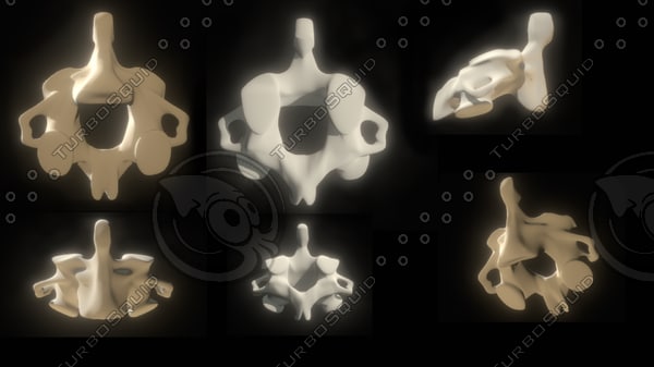 After a successful operation, the model was transferred to the University for student training.
Artificial corneas made on the Nano master SMP-III 3D bioprinter
Source: europepmc.org
In South Korea, about 2000 patients are waiting for corneal donation, and the waiting time for surgery is an average of six years. For patients who cannot find a suitable donor, it is possible to implant artificial corneas consisting of recombinant collagen and synthetic polymers. Unfortunately, they often do not take root and are not completely transparent. This is due to the special structure of the cornea in the form of lattice collagen fibrils, which has not yet been able to be reproduced. A team of researchers from Pohang University of Science and Technology and Kungpuk National University School of Medicine in South Korea have developed a method to 3D print an artificial cornea using patient tissue material.
Source: ithl.co.kr
3D bioprinter with Nano master SMP-III microextrusion system, Musashi Engineering, Tokyo, Japan, with the following parameters:
-
print speed 130mm/min;
-
extrusion speed 0.0024 mm/s;
-
nozzle diameter 0.29 mm;
-
print temperature 4 °C.
The printed and biomaterial-filled cornea was then cultured in an incubator at 37 °C for four weeks.
Source: europepmc.org
A 3D-printed artificial cornea made from decellularized corneal stroma and patient stem cells can completely replace a donor cornea in eye surgery. Since such a cornea is made up of materials derived from the patient's own tissues, it is completely compatible. Cellular 3D printing technology replicates the natural microenvironment of the eye, resulting in transparency similar to that of the human cornea.
Pohang University of Science and Technology Professor Jina Jang said:
"We are confident that this technology will restore vision to many patients suffering from corneal diseases."
Wake Forest Institute for Regenerative Medicine, mobile 3D printer for treating extensive wounds
the place of the damaged. In addition to the fact that this method is additionally traumatic for the victim, in some cases there may not be any healthy skin left on the body for use. Wake Forest School of Medicine has developed a printer that can print skin cells grown from patient tissue directly onto a wound.
Source: 3dnatives.com
The ZScanner Z700 handheld 3D scanner is used to determine the size and depth of a wound. Based on this information, the 3D printer prints subcutaneous, dermal and epidermal skin cells at appropriate depths to completely cover the wound.
Source: 3dnatives.com
The 3D bioprinting system developed by scientists consists of a three-axis moving print head with eight 260 micron diameter nozzles with independent dispensers. Specifically for this device, the researchers created a bioink consisting of autologous dermal fibroblasts and epidermal keratinocytes in a hydrogel carrier.
Bite
Volt Bipolar Surgical Clamp for Laparoscopic Surgery stop bleeding during surgery. It was created for use in minimally invasive (sparing) surgery in 2016 and successfully tested on pig liver.
Source: bitegroup.nl
The design of the device allows easy adjustment of the shaft and tip geometry depending on the patient's anatomy and surgical requirements.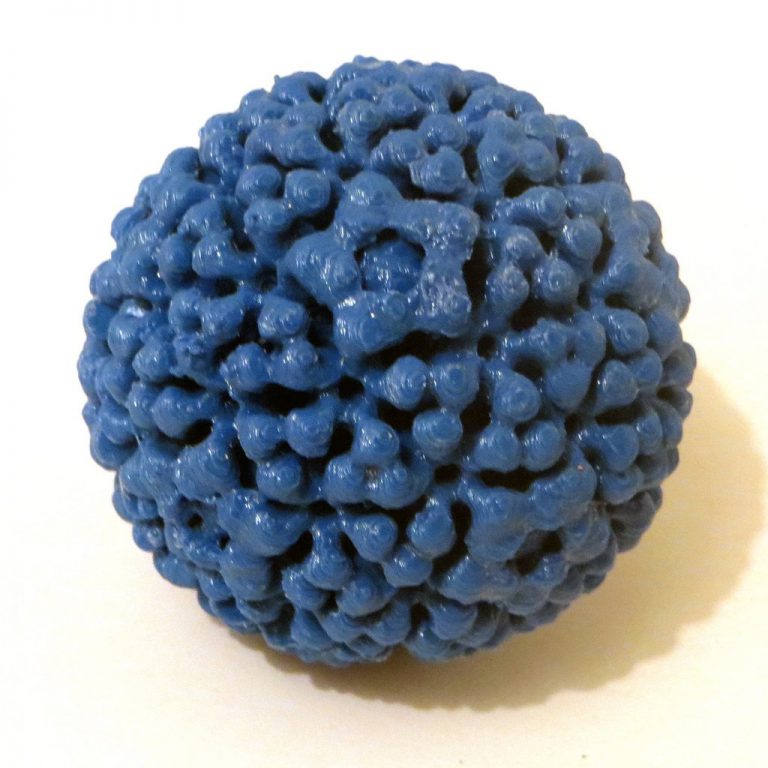 Maneuverable shank - ±65° for lateral movements and ±85° up and down. Flexural stiffness of 4.0 N/mm for connection 1 and 4.4 N/mm for connection 2, significantly higher than previously available guided tools. The tip consists of two 3D printed titanium movable jaws with an opening angle of up to 170°. The instrument is connected to an Erbe electrosurgical unit and is able to successfully coagulate tissue at a temperature of 75 °C, reached in 5 seconds.
Conclusion
Source: intermercados.com.br
The use of additive technologies in medicine is expanding so rapidly that it is more like a revolution in healthcare. The use of 3D printing in medicine enables the individualization of medical devices, medicines and equipment, increases cost efficiency and productivity, reduces waiting times for patients and improves the availability of medical care.
Source
Tags:
3D printing in medicine, dental implants, 3D printing for ears, 3D printed models, Printing surgical instruments, Printing drugs, Prosthetics and dentistry , 3D printer Zortrax M20
More than 10 export/import formats in Renga. Which one to choose?
When working on a building project, you often have to use several programs to solve professional problems.
For the exchange of information with specialized systems Renga supports file formats:
- • DWG, DXF, PDF, OXPS ─ for the exchange of drawings,
- • CSV ─ for exporting parameters, properties and design characteristics,
- • 3DS, LWO, STL, OBJ, COLLADA, FBX, C3D, STEP, IGES, PARASOLID, ACIS, JT and VRML ─ for sharing polygonal and solid models,
- • IFC ─ for exchanging building information models in different views.
While Renga supports import and export to DWG, DXF and PDF files for drawing exchange, as well as OXPS for export only, the choice of file formats for 3D geometry exchange is much larger.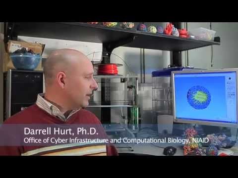
Importing files into a 3D model in Renga
When working with a model in Renga using the Paste from... command, you can import:
- • 2D DWG, DXF and vector PDF. The resulting graphics are converted to Renga objects: model lines, text, and model hatches.
- • 3ds Max 3DS (*.3ds), LightWave (*.lwo), StereoLithography (*.stl), Wavefront object (*.obj), COLLADA (*.dae), Autodesk FBX (*.fbx), and VRML (*.wrl). 3D models in these formats are converted to an Element object. They can be used to create environments and, for example, to transfer a building model from Renga to 3D visualization systems to obtain a photorealistic image. Please note that in the drawings, elements obtained from the listed formats are displayed as a dimensional rectangle. Therefore, if objects must be displayed in drawings, choose other formats.
- • C3D (*.c3d), STEP (*.stp, *.step), IGES (*.igs, *.iges), Parasolid (*.
 x_t, *.x_b), ACIS (*.sat), JT (*.jt). 3D solid models are also converted to an Element object, but their scope is wider, since they are displayed in detail in drawings both in projection and in section, and you can also get Net mass and Net volume for them.
Features of working with 3D formats in Renga
In addition to 3D formats, you can use the IFC4 format to transfer an information model made in other design systems to Renga. An IFC file is opened in Renga without additional settings using the Open... command. The geometric and informational representation of the resulting objects will depend on the source IFC file.
Therefore, when creating an IFC that will later be opened in Renga, you need to pay attention to the settings for exporting to IFC.
Export nothing more, nothing less than the properties you need to work in Renga, because Renga will read all the properties that are in IFC.
IFC files can contain various geometric representations of objects, prepare it according to your needs.
Polygonal or surface model:
- • used for coordination and visualization;
- • allows you to work with properties
Solid model:
- • projected onto the drawing;
- • allows you to work with properties;
- • edited as a Renga model if the build parameters were saved when creating the IFC.
All objects in the IFC file can be managed in the same way as objects created in Renga, for example: copy, move, mirror, assign properties, draw.
The project of an individual residential building. Structural solutions are made in Renga based on IFC. Chief Project Engineer R.V. Mironov
Exporting a 3D model from Renga
The list of 3D formats to which a model can be exported from Renga is slightly different from the import list.
Commands for exporting to various formats are located in the Export menu on the Main Panel. After selecting the Export to 3D command in this menu, 9 formats are available to transfer the model for visualization.0343 OBJ and Collada , for 3D printing ─ STL format, and for exchange with computer-aided design systems C3D, JT, ACIS, STEP and Parasolid .
Let's analyze in more detail which format and in which case it is worth choosing.
When exporting to OBJ and Collada formats, textures applied in Renga are saved next to the model. Thus, models obtained by importing will save all data from the Renga model that can be read by visualization systems.
Lighthouse Sea Museum
Master-Renga 2016 participant. Project of the Lighthouse Sea Museum. Made in Renga, exported in Collada, rendered in Lumion. Author: Ekaterina Kuvshinova
When exporting to the C3D, JT, ACIS, STEP, and Parasolid solid model exchange formats, the structure of the building is preserved, but it may be displayed differently depending on the export format selected.
For example, it is often necessary to transfer a solid model of a building to mechanical engineering CAD to create an environment around equipment, to make measurements. In addition, in such systems, in particular, in KOMPAS-3D, it is possible to obtain a 3D section of the model and its standard projections.
Visualization of industrial site design solutions. The building was created in Renga. Technological equipment was created in KOMPAS 3D. Project authors: ETC NefteGaz Project LLC
Since Renga supports many solid formats, it again becomes a question of choosing the right option for export.
If the model needs to be sent to the customer, select the C3D format, he can evaluate it in the C3D Viewer. Also select C3D if your company uses KOMPAS-3D. And to transfer the model to other design systems, choose the format that is better read by this system. If the system supports all formats, then we recommend choosing JT, as it is the most modern and compact of all.
If you need to transfer the model to be combined with models from other disciplines for visual verification or collision detection, for calculations, for examination, then use the export to the IFC4 format.
IFC is the format with which you can get all the information about the construction object that you could wish for. Therefore, in Renga, export to IFC, unlike export to 3D formats, has a set of settings that allows you to get completely different representations of the model made in Renga, depending on the task.
By default, Renga is configured to export to Reference View, which is intended for:
- • Combining IFC models of various disciplines for visual verification;
- • collision detection;
- • Loading an associated specialist model.
- • calculation of volumes;
- • using the IFC model to link to the construction schedule;
- • Presenting the IFC model to a wider audience.
Learn more