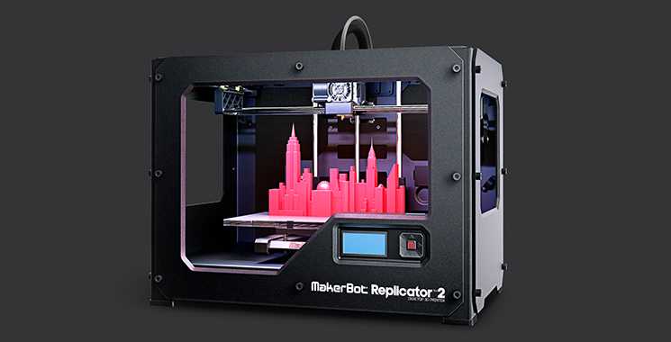
In medicine 3d printing allows more
Contributed: Top 8 healthcare uses for 3D printing
New technology developments have enabled healthcare advances in 3D printing with an estimated $6.08 billion by 2027 in terms of software, hardware, services and materials. The technology has given a boost to customized medicine, allowing a more accurate understanding of patient symptoms and treatment, and generating increased efficiency in the operating room (OR). Advent of 3D printing technology is leaving its mark in specialties such as orthopedics, pediatrics, radiology and oncology, as well as in cardiothoracic and vascular surgery.
Doctors, hospitals and researchers around the world are using 3D printing for:
- preoperative planning and customized surgery.
- medical devices and surgical instruments.
- molds, prostheses and customizable implants.
- 3D digital dentistry and drug administration.
3D printing allows specialists to create reference models using MRI scans and CT in order to help surgeons prepare better for surgeries.
In 2016, a child in Northern Ireland had two unhealed bones injuries his forearm. The child could not rotate his arm more than 50% and was suffering from increased pain. CT scanning and X-rays showed deformed bones, and the treatment required an osteotomy – a four-hour invasive surgery in which the surgeon reshapes the bones to improve rotation. However, the surgeon, printed a 3D model that changed the diagnosis, the surgical intervention and the recovery of the patient:
- It was the tight structures between the bones and not the shape of the bones that limited the child’s rotation ability.
- The procedure was completed in less than 30 minutes, instead of four hours.
- The patient was able to gain 90% arm-range movement four weeks after the intervention.
- The recovery time, the post-operation pain and the scarring decreased considerably.
Such 3D printing is changing preoperative planning which translates into less time spent in the OR, better surgery outcomes for the patients, faster post-op recovery and lower costs for hospitals.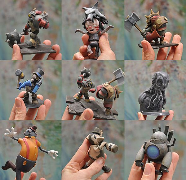
Due to decreased costs of 3D printers and increased availability of CAD/CAM medical software, more hospitals are creating in-house 3D-printed anatomical models. The process entails several steps:
- MRI and CT scans are processed in a stage known as segmentation.
- Each organ and body part type is modeled.
- Models are translated into STL file formats, arranged for printing and sent to the 3D printer.
Rady Children’s Hospital created its own 3D Innovations Laboratory for printing 3D models, including models that mimic human tissue such as airways, hearts and bones. In 2019, the hospital admitted a 7-year-old child born with a single functional heart ventricle (instead of the normal two). The medical team created a 3D-printed model that detailed every vein, artery and valve of the child’s heart, which enabled surgeons to identify the location where blood flow needed rerouting.
Anatomical models that are 3D-printed enable surgeons to plan the operation efficiently and establish better treatment solutions, decrease the operation's duration, and improve research and training for medical students.
3. Designing medical devicesIn order to serve their purpose, medical devices must meet several requirements:
- They need to comprise the perfect balance in terms of size and weight.
- The must match the particular shapes of the human body.
- They have to be functional, and they have to pass specific endurance tests.
Producing medical device to meet these criteria traditionally required extensive time. The alternative found by medical device manufacturers was stereolithography – a process in which a moving laser beam controlled by computer builds the required structure layer by layer. Thus 3D printing has been used to create the prototype of an inhaler, including the needed fixtures and jigs, aiming to:
- Reduce production from one to two weeks to one to two days.

- Reduce cost 90% (from £250 to £11 or $343 to $15).
Customized 3D-printed surgical instruments such as scalpel handles, forceps or clamps help surgeons perform better in the OR, reduce operating time and promote better surgical outcomes for the patients.
Manufactured from materials such as stainless steel, nylon, titanium alloys or nickel, customized surgical instruments are well suited for sterilization. Endocon GmbH – a German medical device producer – has used metal 3D printing to create an alternative surgical tool for hip cup removal. This is traditionally a 30-minute procedure performed with a chisel, but the chisel can sometimes damage the tissue and bones, which results in an uneven surface, making the insertion of a new hip implant difficult.
Endocon’s stainless steel alloy 3D-printed blades, called endoCupcut, enabled precise cutting along the acetabular cup in three minutes and decreased the rejection rate for the replacement, while reducing production time and cost.
While simple prostheses are available in predefined sizes, customized bionic prostheses cost thousands of dollars. This situation affects many children who outgrow their prostheses and need customized replacement parts, which are produced by a handful of manufacturers.
In 2016, Lyman Connor and Eduardo Salcedo created the Lyman’s Mano-matic prosthesis to provide bionic prosthetics to those who need them and cannot afford them. Globally, prostheses designers can use 3D printing to overcome the financial obstacles and time line constraints entailed by this process. The costs of this manufacturing method are significantly lower than traditional methods, and the prostheses are ready in approximately two weeks, making 3D printing a viable solution for customized bionic devices that replicate a human limb's motions and grips.
6. 3D-printed implantsMetal 3D printing enables medical devices designers to produce implants that perform better, match better and last longer, for knees, spine, skull or hips.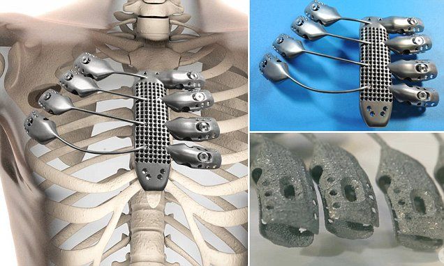
Electron Beam Melting (EBM) is a technology that melts a metal powder layer by layer with the help of an electron beam, thus generating high-accuracy parts. These orthopedic implants provide spongy structures that mimic regular bone tissue, resulting in a higher percentage of osseointegration – the in-growth of a bone into a metal implant.
In 2016, a patient suffering from a tumor that eroded five of his vertebrae was admitted to Peking University Third Hospital. The tumor, caused by a rare form of malignant chordoma, could be removed only through surgery. However, the healing of the extended size bones defects might not have been completely and correctly possible once the lesion would have been removed.
To address this challenge, researchers designed five artificial vertebrae similar to the body structure of the patient using EBM technology. The prosthesis enabled increased stability of the spine, reducing pain and increasing the durability of the device, which allowed the patient to walk without braces two months after the surgical intervention.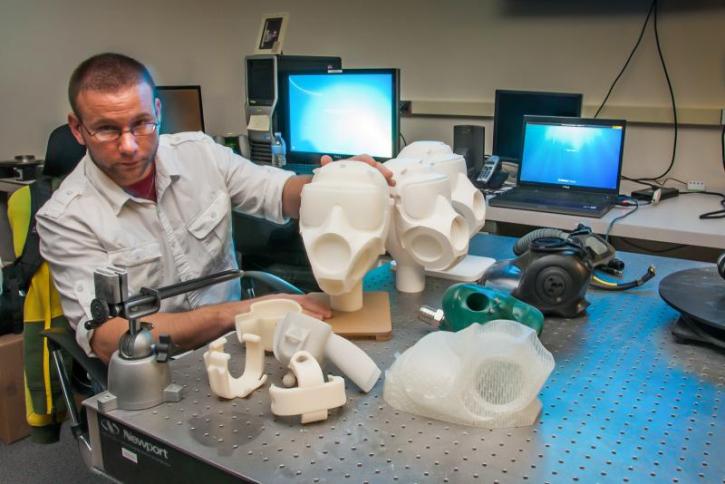
Customized 3D printed implants represent a flexible solution for difficult orthopedic cases and may generate more treatment opportunities in the future.
7. 3D Digital DentistryA recent report indicates that, by 2022, cumulative manufacturing will produce around 500 million dentistry devices and restorations every year, with an estimated $9 billion for the entire dental segment by 2028.
In the dental industry, 3D printing is used for the manufacturing of dentures, surgical guides, bridge models and, most of all, for clear aligners – invisible devices that straighten teeth.
Compared to metal braces, clear aligners are actually invisible and can be taken off when the wearers need to brush their teeth or eat. The traditional production method of clear aligners is a combination of manual and milling processes that requires time and effort. The 3D printing technique speeds up the process, since customized molds for clear aligners can be manufactured directly from digital scans of patients.
Looking for cost-efficient solutions, one dental start-up has perfected an easy process to produce molds for clear aligners:
- Customers take impressions of their teeth with an at-home impression kit or an intraoral scan at a specialized center.
- Impressions and scans are checked by a dental professional, who creates a plan for treatment.
- The company then sends the 3D-printed aligners to the customers.
Therefore 3D printing is a cost-effective method to produce clear aligners, since the setup and tools are not expensive, and their customization is, as proven, direct and simple.
8. Streamlining drug administration3D printing can also simplify drug administration with the help of 3D-printed pills. Polypill is a concept designed for patients suffering from several affections, containing five different drugs compartments and two separate release profiles.
Patients affected by several health issues often take their medication at different hours within the day, and this can be confusing in setting a schedule. This 3D printed pill handles both medication dosage and potential interactions between drugs treating different conditions, so eliminating the need for this scheduling and close monitoring.
The administration of a single customized pill to treat several ailments has multiple advantages:
- increased medication adherence to prescribed treatments.
- customized medication or drug combinations.
- lower production costs, due to the ability to treat more affections at the same time.
- greater accessibility in developing countries to affordable and efficient drugs.
As the price of high-performance 3D printers decreases, more medical professionals use 3D printing to produce cost-efficient customized devices in short periods of time, to design patient-tailored anatomical models, to identify revolutionizing clinical solutions and to create new treatments adapted to patients’ needs.
The advances in 3D-printing technology will attract more customized care and more high-precision medical instruments. At the same time, 3D printing is expected to make an impact in other medical specialties such as ophthalmology, regenerative medicine and bio-printing.
About the Author
Dr. Liz Kwo a serial healthcare entrepreneur, physician and Harvard Medical School faculty lecturer. She received an MD from Harvard Medical School, an MBA from Harvard Business School and an MPH from the Harvard T.H. Chan School of Public Health.
5 Benefits of 3D Printing in Medicine
3D printing is revolutionizing and enhancing various industries and the medical industry is no exception. There are numerous benefits 3D printing provides for the field in order to improve and save patient’s lives.
5 Benefits of 3D printing in the medical industry
1. Complex operations
3D printing plays an important role in training future doctors and preparing for actual operations.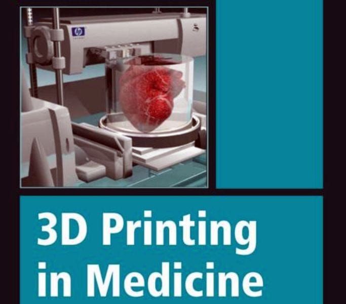 2D images are useful; however, they provide little visualization and do not represent an actual human part. 3D printing, on the other hand, provides models that look realistic and mimic actual human parts. This makes the operational process more accurate and effective.
2. Advanced technology
Future doctors can practice on 3D printed organs. This is much more accurate than for example training on animal organs. Training on human-like, 3D printed parts increases the quality of skills doctors obtain during training and the medical treatment of patients.
3. Intricate care
3D printers create low-cost prosthetics where people need them, for example in war-torn countries. They are an affordable solution for people who cannot afford to buy a prosthetic. Low-cost medical equipment is also important in poverty-stricken countries and remote areas. There are areas where road infrastructure is too bad to deliver medical equipment.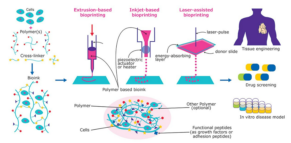 3D printing makes it easier to print the necessary equipment in those villages without having to regularly transport them.
4. Expensive procedures and long waiting time
3D printing allows to 3D print medical and lab equipment. It is possible to 3D print plastic parts of the equipment. This drastically reduces costs and time spent waiting to receive a new medical device from external suppliers. Furthermore, the manufacturing process and further applications are also easier. This makes equipment more readily available and allows low-income or hard to access areas to get 3D printed medical equipment more easily.
5. Customization
Making prosthetics the traditional way is very expensive because they have to be personalized to the individual. 3D printers give users the freedom to choose, e.g. different designs, forms, sizes and colors of their prostheses. This makes every 3D printed piece personalized.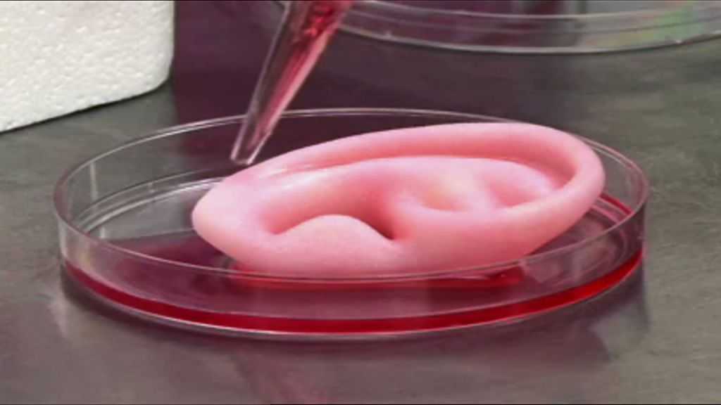 3D printers also allow prosthetics to be more widely available at a lower price.
Do you want to know how you can use 3D printing in your work? CONTACT US and receive a FREE, PERSONALIZED consultation about your possible 3D print solution!
3D PRINTER BUILT FOR PRECISION
The Bolt PRO provides the following benefits to the medical industry.
a. Variety of materials
With the Bolt PRO, you can 3D print with different materials. It is possible to 3D print soft padding where the patient’s bone touches the prosthetic. This increases patient’s comfort and reduces the impact and risk of injury.
b. Large print volume
The Bolt PRO has a large build volume which is great for 3D printing large bones. This allows doctors to have a tangible model they can touch and practice on and better understand the operation.
c. High-temperature hot ends
Bolt PRO enables you to print high-temperature materials that can be sterilized.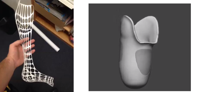 Polypropylene is a great material for medical applications because it offers high levels of heat and is resistant to chemicals.
d. HEPA filter
The Bolt PRO is suitable for the office environment because of its HEPA filter. The filter reduces any negative fumes by 99.9%. This means you can easily have the Bolt PRO at your office and use it for printing on the spot.
Are you interested in a success story of Mackay Hospital using 3D printing to enhance their surgeon’s skills? Click HERE.
3D printing applications in medicine will keep on improving and increasing people’s quality of life. We are certain that in the end, 3D printers will be a vital part of medical processes.
5 Innovative Medical Applications for 3D Printing
Personalized and precise medical solutions are gaining popularity. New tools and advanced technologies bring doctors closer to patients by providing treatments and devices that meet the needs of each individual.
The expansion of 3D printing technology in healthcare has made a huge contribution to improving the quality of medical services. With new tools and treatment approaches developed using 3D printing, patients feel that their treatment becomes more comfortable and personal. For physicians, the new technology available allows them to better analyze complex cases and provides new tools that can ultimately raise standards of care.
Later in this article, you'll learn about five areas, from models for surgical planning to vascular systems and bioreactors, in which 3D printing is used in healthcare, and why many healthcare professionals see great potential in this technology.
In today's medical practice, 3D printed anatomical models based on patient body scans are becoming more indispensable tools, as they provide more personalized and accurate treatment. As cases become more complex and standard case times become more important, visual and tactile anatomical models are helping surgeons to better understand their task, communicate more effectively, and communicate with patients more easily.
Medical professionals, hospitals and research institutions around the world use 3D printed anatomical models as a reference tool for preoperative planning, intraoperative imaging, and for sizing medical instruments or presetting equipment for both standard and very complex procedures, which is reflected in hundreds of scientific publications.
3D printing makes 3D printing affordable and easy to create customized patient anatomical models based on CT and MRI data. The peer-reviewed scientific literature demonstrates that they help clinicians better prepare for surgery, resulting in significant cost and time savings. At the same time, patient satisfaction is also increased through reduced anxiety and reduced recovery time.
Physicians can use individual patient anatomical models to explain the procedure to the patient, making it easier to obtain patient consent and reduce patient anxiety.
Preparation for surgery using preoperative models can also affect the effectiveness of the treatment.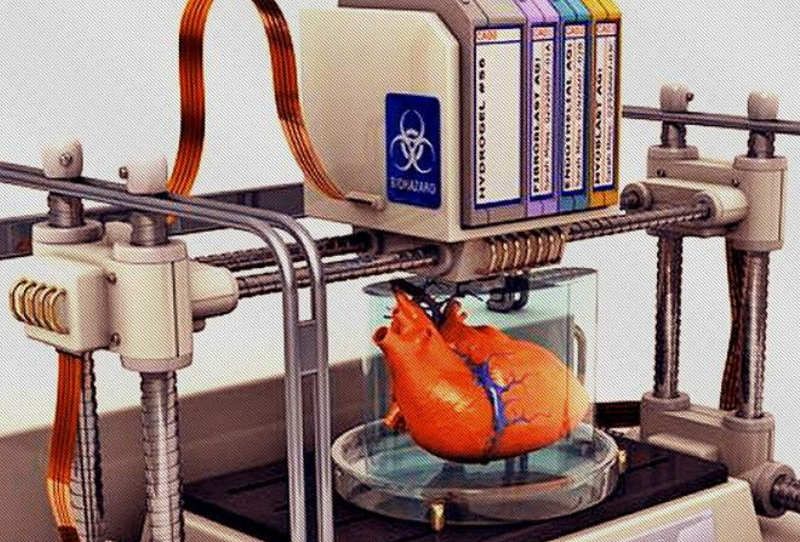 The experience of Dr. Michael Ames confirms this. After obtaining bone replications from the young patient's forearm, Dr. Ames realized that the injury was different from what he expected.
Based on this information, Dr. Ames chose a new soft tissue procedure that was much less invasive, reduced downtime, and resulted in much less scarring. Using imprinted bone replication, Dr. Ames explained the procedure to the young patient and his parents and obtained their consent.
Physicians can use patient-specific surgical models to explain the procedure beforehand, improving patient consent and lowering anxiety.
Result? The operation lasted less than 30 minutes instead of the originally planned three hours. With this reduction in operating time, the hospital avoided a cost of about $5,500 and the patient recovered faster.
According to Dr. Alexis Dang, Orthopedic Surgeon at UC San Francisco and Veterans Affairs Medical Center San Francisco: “All of our full-time orthopedic surgeons and nearly all of our full-time surgeons part-time, used 3D printed models to treat patients at a Veterans Medical Center in San Francisco.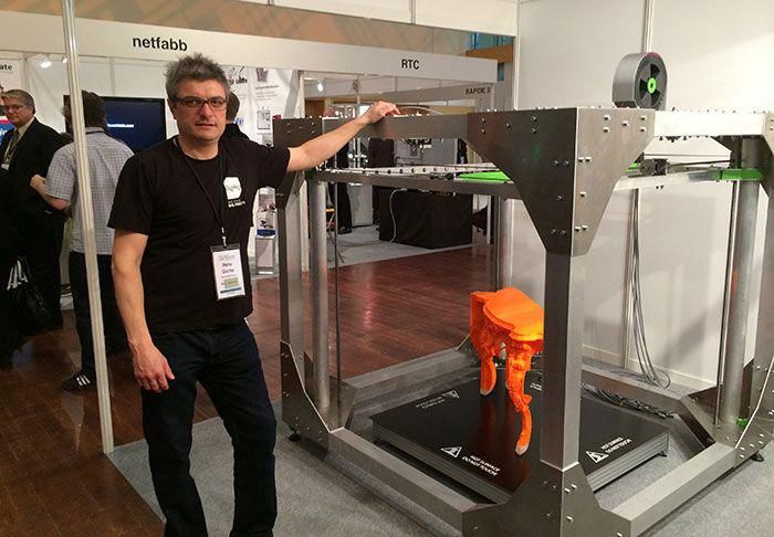 We could all see that 3D printing improves the efficiency of our work.”
The advent of new biocompatible medical polymers for 3D printing has opened up opportunities for the development of new surgical instruments and techniques to further improve clinical operating procedures. These include sterilizable trays, contoured surgical guides, and implant models that can be used to determine the size of an implant prior to surgery, helping surgeons reduce time and improve accuracy in complex procedures.
Anatomical model of a hand with elastic resin skin for 3D printing.
Todd Goldstein, PhD, lecturer at the Feinstein Institute for Medical Research, is unequivocal about the importance of 3D printing technology to the work of his department. He estimates that if Northwell used 3D-printed models 10-15% of the time, it could save $1,750,000 a year.
“Whether it's prototyping medical devices, complex anatomical models for our children's hospital, designing training systems, or making surgical templates for dental clinics, [3D printing technology] has increased our capabilities and reduced our costs in a variety of areas.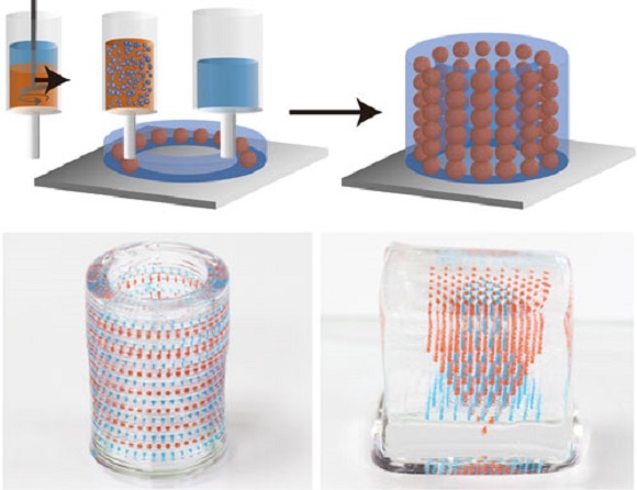 In doing so, we were able to produce instruments for treating patients that would be almost impossible to recreate without our sought-after stereolithography 3D printer,” says Goldstein.
3D printing has become virtually synonymous with rapid prototyping. The ease of use and low cost of 3D printing in-house has also revolutionized product development, with many medical instrument manufacturers adapting the technology to produce entirely new medical devices and surgical instruments.
Over 90 percent of the top 50 medical device companies use 3D printing to create accurate medical device prototypes and fixtures and fittings to simplify testing.
According to Alex Drew, Principal Mechanical Engineer at DJO Surgical, an international medical device supplier, “Before DJO Surgical purchased [Formlabs' 3D printer], we printed nearly all of our prototypes outsourced. Today we are working with four Formlabs printers and are very pleased with the results. The speed of 3D printing has doubled, the cost has been reduced by 70%, and the level of detail allows you to effectively coordinate designs with orthopedic surgeons.
Medical companies such as Coalesce are using 3D printing to create accurate medical device prototypes.
3D printing helps speed up the design process by allowing complex designs to be iterated over in days instead of weeks. When Coalesce was tasked with building an inhaler device that could digitally evaluate an asthma patient's inspiratory flow profile, outsourcing would result in a significant increase in production time for each prototype. Before sending the project files to a third party company for the physical implementation of the project, they would have to be carefully developed and carried through various iterations.
Instead, desktop stereolithographic 3D printing allowed Coalesce to handle the entire prototyping process in-house. The prototypes were suitable for use in clinical trials and looked just like the finished product. Moreover, when the company demonstrated the device, its customers mistook the prototype for the final product.
Overall, the introduction of in-house manufacturing resulted in an exceptional reduction in prototyping time by 80–90%.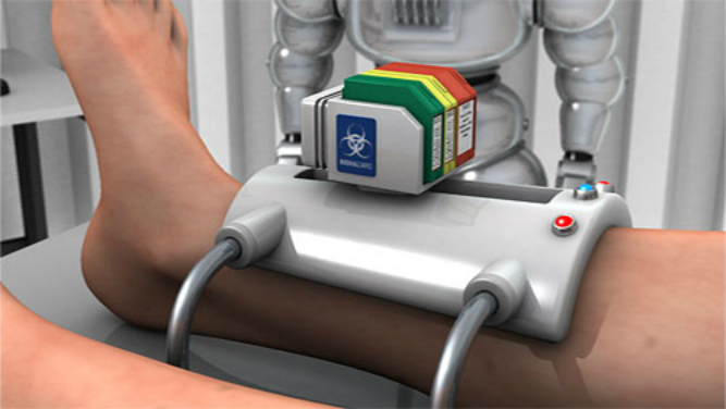 In addition, the models took only eight hours to print and were finished and painted in a matter of days, while outsourcing the same process would take a week or two.
Hundreds of thousands of people lose limbs every year, but only a fraction of them are able to restore limb function with a prosthesis.
Conventional dentures are only available in a few sizes, so patients must adjust to what fits best. On the other hand, custom bionic prostheses that mimic the movements and grips of a real limb based on the impulses of the surviving limb muscles are so expensive that they can only be used by patients living in developed countries with the best medical insurance. In the case of children's prostheses, the situation is aggravated even more. Children grow up and inevitably outgrow their prostheses, which, as a result, require costly modifications.
The difficulty lies in the lack of manufacturing processes that would allow for individual orders at an affordable price. But increasingly, prosthetists are looking to reduce these high financial barriers to rehabilitation with the flexible design capabilities of 3D printing.
Initiatives like e-NABLE allow people around the world to learn about the possibilities of 3D printed prostheses. They are driving an independent movement in the prosthesis industry by offering information and free open source projects so that patients can get a custom-designed prosthesis for as little as $50.
Other inventors, such as Lyman Connor, go even further. With only a small fleet of four desktop 3D printers, Lyman was able to fabricate and customize his first mass-produced prostheses. His ultimate goal? Create a customizable fully bionic arm that will cost incomparably less than similar prostheses that retail for tens of thousands of dollars.
Researchers at the Massachusetts Institute of Technology have also found that 3D printing is the best method for making more comfortable prosthetic sockets.
In addition, the low cost of manufacturing these prostheses, as well as the freedom that comes with being able to design custom designs, speak for themselves. 3D printed prostheses have a lead time of just two weeks, and then they can be tried and serviced at a much lower cost than traditional counterparts.
As costs continue to fall and material properties improve, the role of 3D printing in healthcare will no doubt become more important.
The same high financial barriers that are seen in prosthetics are common in orthoses and insoles. Like many other patient-specific medical devices, custom-made orthoses are often not available due to their high cost and take weeks or months to manufacture. 3D printing solves this problem.
Confirmation is the example of Matej and his son Nick. Nick was born in 2011. Complications during preterm birth led to the fact that he developed cerebral palsy, a pathology that affects nearly twenty million people worldwide. Matei was delighted with how determined his son was to overcome the limitations of his illness, but he was faced with a choice between a standard, off-the-shelf orthosis that would be uncomfortable for his son, or an expensive custom solution that would take weeks or months to manufacture and ship.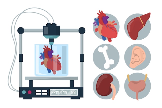 , and from which the child would quickly grow.
He decided to take matters into his own hands and began to look for new ways to achieve his goal. Thanks to the opportunities provided by digital technologies, in particular 3D scanning and 3D printing, Matei and Nika's physiotherapists were able to develop a completely new innovative workflow for the manufacture of ankle orthoses through experiments.
The resulting 3D-printed, custom-fit orthosis that provides support, comfort, and motion correction helped Nick take his first steps on his own. This non-standard orthopedic device reproduced the functionality of the highest-class orthopedic products, at the same time it cost many times less and did not require any additional settings.
Professionals around the world are using 3D printing as a new method of manufacturing custom insoles and orthoses for patients and clients, as well as a range of other physiotherapy tools. In the past, undergoing a course of physiotherapy with the use of individual physiotherapy instruments carried many difficulties.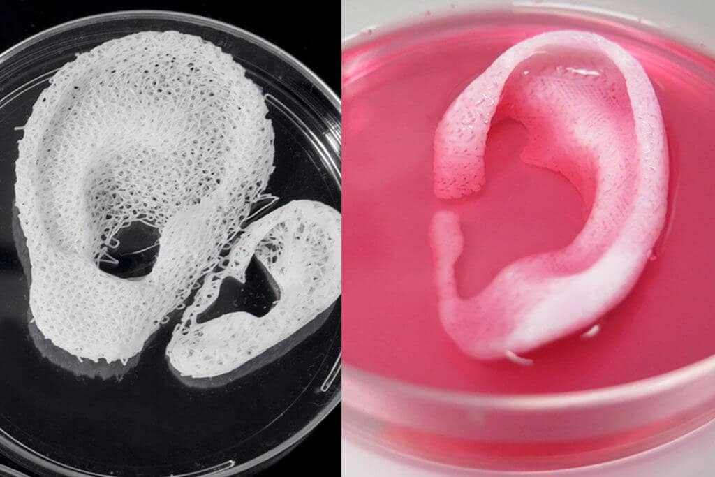 Often there was a situation when patients had to wait a long time for a finished product, which at the same time did not provide proper comfort. 3D printing is step by step changing this status quo. Data confirms that 3D printed insoles and orthoses offer a more precise fit and lead to better therapeutic outcomes, which means greater comfort and benefit for patients.
The usual treatments for patients with severe organ damage today are autografts, transplantation of tissue from one area of the body to another, or transplantation of a donor organ. Researchers in bioprinting and tissue engineering hope to expand this list soon with on-demand fabrication of tissues, blood vessels, and organs.
3D bioprinting is an additive manufacturing process that uses materials known as bioink (a combination of living cells and a compatible substrate) to create tissue-like structures that can be used in medicine. Tissue engineering combines new technologies, including bioprinting, which make it possible to grow replacement tissues and organs in the laboratory for use in the treatment of injuries and diseases.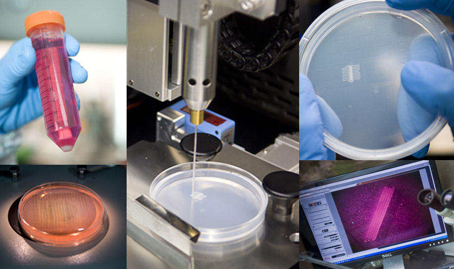
Using high-precision 3D printing, researchers such as Dr. Sam Pashne-Tala from the University of Sheffield are opening up new possibilities for tissue engineering.
In order to direct cell growth to form the necessary tissue, Dr. Pashne-Tala grows living cells on a laboratory scaffold that provides a template of the required shape, size and geometry. For example, to create a blood vessel for a patient with cardiovascular disease, a tubular structure is needed. The cells will multiply and cover the scaffold, taking on its shape. Then the scaffold is gradually destroyed, and the living cells take the form of the target tissue, which is cultured in a bioreactor - a chamber that contains the cultured tissue and can reproduce the internal environment of the body so that the cultured tissue acquires the mechanical and biological characteristics of organic tissue.
3D printed bioreactor chamber with tissue engineered aorta miniature inside. The tissue is cultured in a bioreactor to acquire the mechanical and biological characteristics of the organic tissue.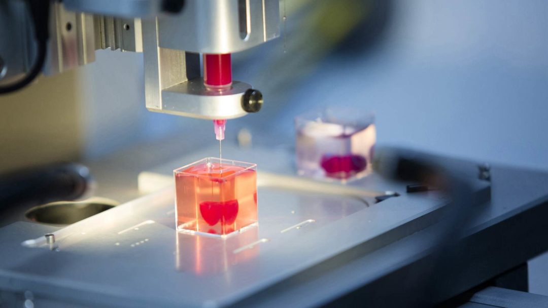
3D printed bioreactor chamber with tissue engineered aorta miniature inside. The tissue is cultured in a bioreactor to acquire the mechanical and biological characteristics of the organic tissue.
This will allow scientists to design patient-specific vascular grafts, expand surgical care, and provide a unique platform for testing new vascular medical devices for people suffering from cardiovascular disease, which is currently the leading cause of death worldwide. The ultimate goal is to create blood vessels that are ready for implantation in patients. Since tissue engineering uses cells taken from a patient in need of treatment, this eliminates the possibility of rejection by the immune system, which is the main problem of modern transplantology.
3D printing has proven its ability to solve the problems that exist in the production of synthetic blood vessels, in particular, the difficulty of recreating the required accuracy of the shape, size and geometry of the vessel. The ability of printed solutions to clearly reflect the specific characteristics of patients was a step forward.
According to Dr. Pashne-Tal: “[Creating blood vessels using 3D printing] makes it possible to expand the possibilities of surgical care and even create designs of blood vessels for a specific patient. Without the existence of high-precision affordable 3D printing, the creation of such forms would not be possible.”
We are witnessing significant advances in the development of biological materials that can be used in 3D printers. Scientists are developing new hydrogel materials that have the same consistency as organ tissues present in the human brain and lungs, which can be used in a range of 3D printing processes. Scientists hope that they will be able to implant them into the body as a "scaffold" for cell growth.
Although bioprinting of fully functional internal organs such as the heart, kidneys and liver still looks futuristic, hybrid 3D printing at very high speed opens up more and more new horizons..jpg)
It is expected that sooner or later the creation of biological matter on laboratory printers will lead to the generation of new, fully functional 3D printed organs. In April 2019, scientists at Tel Aviv University 3D-printed the first heart using biological tissue from a patient. A tiny copy was created using the patient's own biological tissues, which made it possible to fully match the immunological, cellular, biochemical and anatomical profile of the patient.
“At this stage, the heart we printed is small, about the size of a rabbit heart, but normal-sized human hearts require the same technology,” says Prof. Tal Dvir.
The first 3D bioprinted heart created at Tel Aviv University.
Precise and affordable 3D printing processes, such as desktop stereolithography, are democratizing access to technology, enabling healthcare professionals to develop new clinical solutions and quickly produce medical devices with individual characteristics, and doctors around the world to offer new types of therapy.
As 3D printing technologies and materials improve, it will continue to expand personalized treatment and deliver high performance medical devices.
Learn more about 3D printing applications in healthcare
Overview of 3D printing applications in medicine
Use of 3D printing in medicine
Source: docwirenews.com
3D printing has been used in medicine since the early 2000s, when this technology was first used to make dental implants. Since then, the use of 3D printing in medicine has expanded significantly, with doctors around the world describing ways to use 3D printing to produce ears, skeletal parts, airways, jawbones, eye parts, cell cultures, stem cells, blood vessels and vasculature, tissues and organs, new dosage forms and much more.
Source: zortrax.com
The use of files with models for 3D printing provides an opportunity for the exchange of work among researchers. Instead of trying to reproduce the parameters described in scientific journals, doctors can use and modify ready-made 3D models. To this end, in 2014, the National Institutes of Health established the 3dprint.nih.gov exchange to facilitate the exchange of open source 3D models for medical and anatomical devices, custom equipment, and mock-ups of proteins, viruses, and bacteria.
Source: 3dprint.com
Modern medical use of 3D printing can be divided into several broad categories: tissue and organ fabrication, prostheses, implants and anatomical models, instrument printing, and pharmaceutical research.
Top five uses for 3D printing in medicine
Preparation for operations and student education
Source: 3dprint.com
Taking into account individual differences and features of the anatomy of a particular human body, 3D printed models can be used to prepare surgical operations.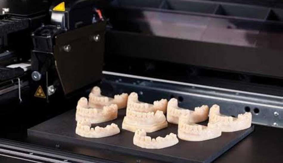 Having a doctor have a tangible model of a particular patient's organ, made, for example, based on the results of CT (computed tomography) for study or to simulate an operation, significantly reduces the risk of medical errors.
Source: openbiomedical.org
The use of 3D models for training surgeons and students is preferable to training on cadavers, since it does not create problems in terms of availability and cost of objects. Cadavers often lack appropriate pathology, so they are more suitable for anatomy lessons than for presenting a patient with a disorder appropriate to the topic under study. With the help of 3D printing, it is possible to create a model of any organ with any known pathology.
Source: ncbi.nlm.nih.gov
two-dimensional images.
Bioprinting of tissues and organs
Source: hbr.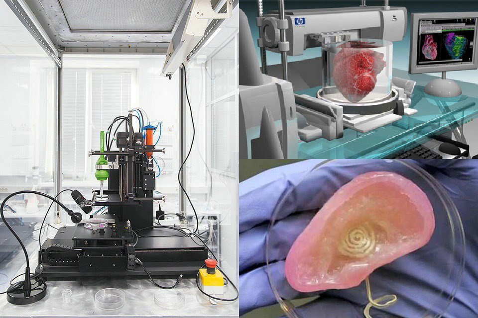 org
org
Bioprinting is one of the many types of 3D printing used in the medical field. Instead of printing using plastic or metal, bioprinters use a syringe dispenser to apply bioink (layers of living cells or a structuring base for them) to create artificial living tissue. In addition to being used as an alternative to donor tissues, such tissue constructs or organoids can be used for medical research.
Source: press.ginkgo3d.com
Although 3D bioprinting systems can be laser, inkjet, or extrusion, inkjet bioprinting is the most common. Multiple printheads can be used to accommodate different types of cells (organ-specific, blood vessel cells, muscle tissue), which is a major challenge in the fabrication of heterocellular tissues and organs. 3D printing with biological materials can be used to regenerate tissues, and in the future, organs, directly on the patient.
Printing Surgical Instruments
Volt Grip Details, Source: bitegroup.nl
Today's surgeons are trying to perform operations with as little trauma to the patient as possible, so they very often require a personalized tool. The use of 3D printing makes it possible to create such tools within hours.
Volt capture model visualization, Source: bitegroup.nl
Now the doctor can independently modify the finished model, giving it the necessary size and shape for convenience and efficiency. Dentists can now create, for example, individual guides right in front of the patient, eliminating the possibility of damage to healthy teeth during prosthetics.
About the clamp Volt, from the photos above, read further in the section “Examples of use”.
Here's how students at Duke University in Durham, North Carolina create tools using metal 3D printing.
"Printing" drugs
Source: mdpi.com
3D printing technologies are already being used in pharmaceutical research and personalized medicine, and their scope is constantly expanding. 3D printing enables precise dose control of drugs and the production of dosage forms with complex drug release profiles and prolonged action. Now pharmacists can analyze a patient's pharmacogenetic profile and other characteristics such as age, weight, or gender to determine the optimal dose and sequence of medications. If necessary, the dose may be adjusted, depending on the clinical response. With 3D printing, it is possible to produce personalized medicines in completely new formulations, such as tablets containing multiple active ingredients, either as a single mixture or as complex multi-layered tablets.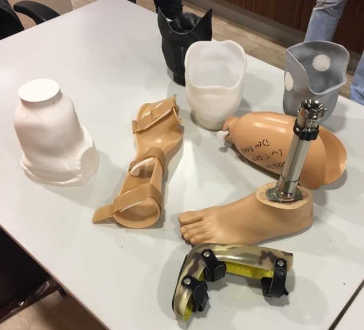
Prosthetics and Dentistry
Source: eos.info
3D printing has been successfully used in medicine for the manufacture of complex custom prostheses or surgical implants. Implants and prostheses of any possible geometry can be made by converting X-ray, MRI or CT images into a 3D printable model using special software.
The rapid fabrication of custom implants and prostheses solves a pressing problem in orthopedics where standard implants often do not fit the patient. This is also true in neurosurgery: skulls are individually shaped, so it is difficult to standardize a cranial implant. Previously, surgeons had to use various tools to modify and fit implants, sometimes right during the operation. The use of 3D printers makes this procedure unnecessary. Additive technologies are especially in demand when it is necessary to urgently manufacture implants.
A real revolution in dentistry occurred with the advent of 3D technologies.
Source: hypowerfuel.com
First, complete and accurate 3D scanning of the oral cavity is now possible. Secondly, the use of 3D printing has made it possible to create prostheses that absolutely fit the anatomy of the patient, without the need for a long and unpleasant fit. The radical reduction in the share of manual labor in the manufacture of prostheses or veneers has reduced the required tolerances in production, expanded the list of materials used and increased patient satisfaction with the results of the doctor's work.
Application examples
Printing a model of the heart of a four-year-old patient, Zortrax M200 3D printer
In the photo: the assembled heart model. Source: zortrax.com
At the Medical University of Gdansk (Poland) to prepare for an operation to treat a complex congenital heart disease (tetrads of Fallot - abnormal functioning of the pulmonary artery heart valve) in a four-year-old patient, specialists from the Department of Pediatric Cardiology and Congenital Heart Diseases , together with colleagues from the Department of Cardiac Surgery and Radiology, used the Zortrax M200 3D printer.
Photo: artificial pulmonary valve. Source: zortrax.com
The modern method of treatment consists in inserting a catheter through the femoral vein, through which an artificial valve is fed to the heart for implantation. This is a very complex operation that requires the doctor to have detailed knowledge of the individual characteristics of the patient's anatomy.
In the photo: a heart model during printing. Source: zortrax.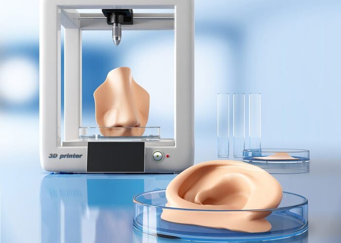 com
com
Until now, doctors could only rely on a 3D model on a computer screen created from CT and MRI images, and such a reconstruction is not always enough to get a complete picture of the real organ and possible complications .
Source: zortrax.com
Having a highly detailed tactile model of a patient's living organ in preparation for surgery can be critical to its success. Even experienced surgeons have appreciated the potential of the new technology. Previously, it was difficult to notice individual features and deformations, now it has become tangible and accessible for closer study.
The model was printed within 24 hours. The Z-ULTRAT material was used to print the heart, and the Z-GLASS material was used to print the vessels. After a successful operation, the model was transferred to the University for student training.
Artificial corneas made on the Nano master SMP-III 3D bioprinter
Source: europepmc. org
org
In South Korea, about 2000 patients are waiting for corneal donation, and the waiting time for surgery is an average of six years. For patients who cannot find a suitable donor, it is possible to implant artificial corneas consisting of recombinant collagen and synthetic polymers. Unfortunately, they often do not take root and are not completely transparent. This is due to the special structure of the cornea in the form of lattice collagen fibrils, which has not yet been able to be reproduced. A team of researchers from Pohang University of Science and Technology and Kungpuk National University School of Medicine in South Korea have developed a method to 3D print an artificial cornea using patient tissue material.
Source: ithl.co.kr
3D bioprinter with Nano master SMP-III microextrusion system, Musashi Engineering, Tokyo, Japan, with the following parameters:
-
print speed 130mm/min;
-
extrusion speed 0.
 0024 mm/s;
 -
nozzle diameter 0.29 mm;
-
print temperature 4 °C.
The printed and biofilled cornea was then cultured in an incubator at 37°C for four weeks.
Source: europepmc.org
A 3D-printed artificial cornea made from decellularized corneal stroma and patient stem cells could completely replace a donor cornea in eye surgery. Since such a cornea is made up of materials derived from the patient's own tissues, it is completely compatible. Cellular 3D printing technology replicates the natural microenvironment of the eye, resulting in transparency similar to that of the human cornea.
Pohang University of Science and Technology Professor Jina Jang said:
"We are confident that this technology will restore vision to many patients suffering from corneal diseases.:quality(80)/images.vogel.de/vogelonline/bdb/1612700/1612740/original.jpg) "
"
Wake Forest Institute for Regenerative Medicine Mobile 3D printer for treating extensive wounds
the place of the damaged. In addition to the fact that this method is additionally traumatic for the victim, in some cases there may not be any healthy skin left on the body for use. Wake Forest School of Medicine has developed a printer that can print skin cells grown from patient tissue directly onto a wound.
Source: 3dnatives.com
The ZScanner Z700 handheld 3D scanner is used to determine the size and depth of a wound. Based on this information, the 3D printer prints subcutaneous, dermal and epidermal skin cells at appropriate depths to completely cover the wound.
Source: 3dnatives.com
The 3D bioprinting system developed by scientists consists of a three-axis moving print head with eight 260 micron diameter nozzles with independent dispensers. Specifically for this device, the researchers created a bioink consisting of autologous dermal fibroblasts and epidermal keratinocytes in a hydrogel carrier.
Bite
Volt Bipolar Surgical Clamp for Laparoscopic Surgery stop bleeding during surgery. It was created for use in minimally invasive (sparing) surgery in 2016 and successfully tested on pig liver.
Source: bitegroup.nl
The design of the device allows easy adjustment of the shaft and tip geometry depending on the patient's anatomy and surgical requirements. Maneuverable shank - ±65° for lateral movements and ±85° up and down. Flexural stiffness of 4.0 N/mm for connection 1 and 4.4 N/mm for connection 2, significantly higher than previously available guided tools. The tip consists of two 3D printed titanium movable jaws with an opening angle of up to 170°. The instrument is connected to an Erbe electrosurgical unit and is able to successfully coagulate tissue at a temperature of 75 °C, reached in 5 seconds.![]()


