3D scanner medical
The revolution in medical applications of 3D scanning
Posted By Kat Plewa on Feb 27, 2019 |
We talk a lot about 3D printing in the medical industry, from 3D printed knee replacements to hearts. But there is much more to discover within 3D technologies. For instance, 3D scanning. This technique enables doctors around the world to literally look inside the patient in just a matter of seconds! What are other benefits of 3D scanning for medical applications? Let’s find out!
How is 3D scanning beneficial for the medical industry?
3D scanning gives doctors an exact image of their patient and allows them to really study different solutions without testing them on the patient directly. With this technology, they are able to visualize inside a patient’s body. No other technology has ever given doctors such a close look inside their patients. 3D scanners can also be portable and are always very fast, which sometimes is a matter of life and death for the injured. Such an ambitious project started father and son, after years of development, their impressive work resulted in Mars– the 3D scanner which sees through people.
Safer procedure
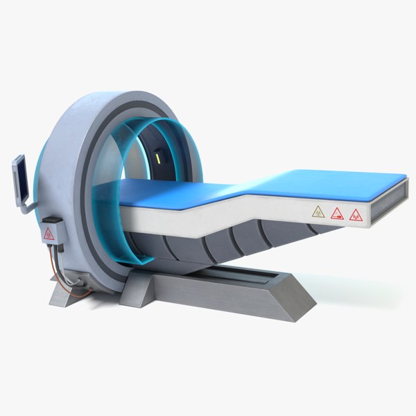
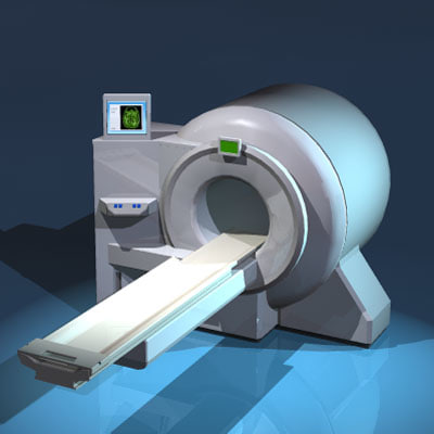
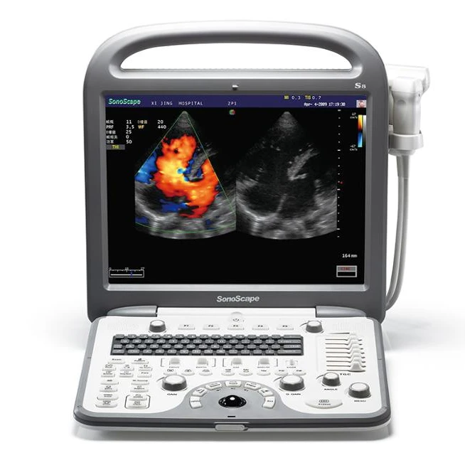
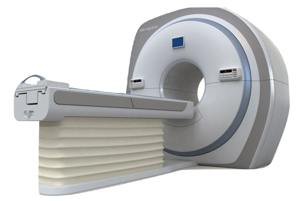
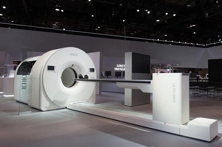
A very important quantity of 3D scanning is that it is absolutely harmless. X-rays and MRIs use radiation to produce pictures of the body which always carries a risk. There is no such risk with a 3D scanner to capture the position of the object in reality and register the data in virtual reality either via photos, light or a laser beam.
https://www.3ders.org/articles/20131030-3d-scanner-maker-creaform-acquired-by-ametek-for-120-million.html
Faster diagnosis
Let’s start with how quick 3D scanning is. When it comes to saving someone’s life every minute counts. Thanks to 3D scanning all the important data about the patient’s condition can be gathered in a matter of seconds and the doctors will have a better view on what should be their next step to save or improve the patient’s life. With 3D scanning, the doctors will be able to diagnose different diseases and disorders at a much earlier stage.
Portable
3D scanners, especially for medical applications, can also be portable. This is especially useful for orthopedics. but also means that doctors around the world can reach more people and therefore provide them with the medical assistance they need.
http://3dprintingireland.com/portfolio/pharmaceutical-medical/attachment/scanmain/
High level of customization
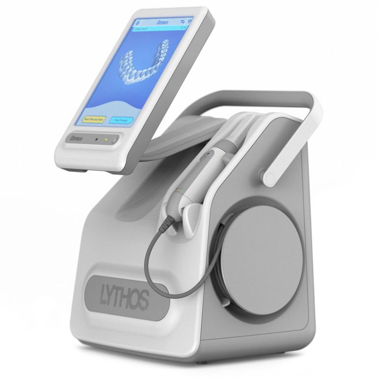
3D scanning increases accuracy, soon we can say goodbye to heavy and uncomfortable plasters. The future of made to measure, highly personalized and much more precise casts is near. Mobile 3D scanners make it very easy to get the shape of the limb and based on that a 3D model is made and then printed. This is also speeding up the whole process.
This aspect of 3D scanning provides a much better fit and is crucial for prosthetic design. Prosthetic legs or arms make it so much easier for the users to carry out everyday tasks.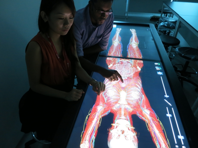 But wearing them is not always easy. They are sometimes heavy and uncomfortable. 3D scanning is the solution to make the prosthesis fit perfectly to the user’s limb. Medical applications of 3D scanning clearly improve this industry.
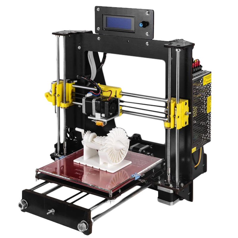
3D printed models
3D scanning combined with 3D printing is extremely important for surgical and dental fields. Those medical applications help as early as in schools, where future doctors can practice and study different medical cases. 3D scanning has also been useful before performing surgeries as doctors can have a true look inside the patient, study tumors or other changes in the body before they begin.
Another medical application of 3D scanners is in the dental industry. Thanks to 3D scans it is so much easier to produce aligners, implants, dentures and crowns which perfect fit within the patient’s jaw. Read more about 3D printing in the dental industry here. And since we are on the topic of jaws created thanks to 3D technologies, let’s see some real-life examples of using the benefits of 3D scanning for medical applications.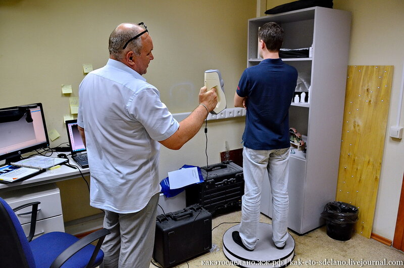
A cancer survivor with a 3D printed jaw
Anelia Myburgh is from Australia and despite her young age, she already has experienced a lot in life. It all began with just a small bump she complained about, but it turned out to be a dangerous cancer tumor. A life-saving surgery left her scared forever. 80% of her upper jaw had to be removed leaving her face disfigured.
That, of course, had a huge impact on her life. Daily tasks such as eating, drinking and talking were not as easy to achieve anymore. On top of that Anelia’s face changed visually and it really brought her confidence down. All she wanted was to just look normal again. “I just want to be able to walk down the street and not have people stare,” Myburgh commented, “That is my ultimate goal.”
https://www.dailymail.co.uk/news/article-6453939/Woman-31-lost-jaw-teeth-cancer-fitted-3D-jaw-world-makeover.html
3D technologies came to rescue. To demonstrate and test different solutions doctors 3D printed an exact model of Anelia’s skull. There were able to take a very close look and study her case in depth to provide her with the best solution. The use of Additive Manufacturing doesn’t finish there; her new jaw implant was in fact also 3D printed! It was a fast and highly accurate way to help the patient’s case by 3D printing a titanium frame for her.
What is next for medical applications of 3D scanning?
3D scanning is already used in medical school, institutions and hospitals. Surely, soon portable 3D scanners will be a standard part of ambulance equipment and paramedics will be able to diagnose the patient even before reaching the hospital with much better accuracy. As you can see, 3D scanning is safe for the patient, quick and helps to bring the comfort of customized aids to save or improve their quality of life.
The advantages of 3D scanning and 3D printing apply not only to the medical industry. High personalization and fast production are very important in many fields. Start improving your manufacturing process today! With online 3D printing service like Sculpteo, it’s as easy as uploading your file. Don’t wait for your competitors to start 3D printing, do it yourself.
Get the best industry insights with our weekly Newsletter and Facebook!
MEDICINE – SMARTTECH 3D scanners
Skip to contentMEDICINEadmin2019-07-22T13:54:36+02:00
Touchless measurement methods can be sufficiently used in medicine. Structured light is currently the safest and the most complex method for wound or skin deviation analysis. Thanks to the possibility of capturing the real color of scanned surface the doctors and researchers gather information of all important areas that can might keep the normal geometry.
3D scanning releases the patient from staying in clinic for to long. Within just few seconds we are able to capture all information about surfaces and colors that can be analyzed further. Patient need to present only during 3D scanning process which is not only rapid but also completely painless.
The lack of X ray and other harmful radiation allows the doctor to work with a 3D scanner without a need of leaving the patient, moreover it’s save for pregnant woman or deeply wounded patients.
The method is is simple – The straight fringes are projected by LED projections systems on the surface that must be measured.
The curvature of the fringes is being captured by the detection camera that sends the digital images to the software. SMARTTECh4Dmeasure software transform the images to the point cloud that presents the geometry and color of measured surface. That’s the way we get the information that can be analyzed in the necessary way – if it’s for plastic surgeries, healing processes monitoring, posture defects analysis etc.
Thanks to ease of usage, SMARTTECh4D scanners can be easily used by the people who are not familiar with metrology. The scanners does not need the difficult calibration process. They are ready to use directly after taking them out from the case. Furthermore – the operating SMARTTECh4Dmeasure software is intuitive and allows to provide 3D scanning as well as automate data analysis.
SMARTTECH 3D scanners are already used in Japanese research institutes such as AIST or UNITIKA where they are used to provide complex measurements on whole human body or it’s parts.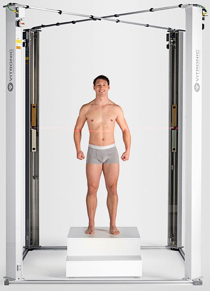 They took part in the European research projects of measurement the sportsman legs.
They were also used to scan Robert Lewandowski to beat the World Guinness Record for the biggest 3D printed statue.
PRODUCTS INTENDED FOR MEDICAL INDUSTRY
SMARTTECh4D
MED
Read more
SMARTTECh4Dmeasure
Read more
SMARTTECh4D
UNIVERSE
Read more
HOW WE HELPED OTHERS FROM YOUR INDUSTRY
admin2019-09-05T15:07:09+02:00
3D technology is revolutionizing the medical industry
The medical industry is currently one of the fastest-growing ones. Almost every day new ideas are born. Unfortunately, the process [...]
admin2019-08-23T10:52:58+02:00
The 3D scanner in a unique design for creating an anatomical baby bottle
Today, the accessory industry for newborns and infants is a dynamically developing business sector.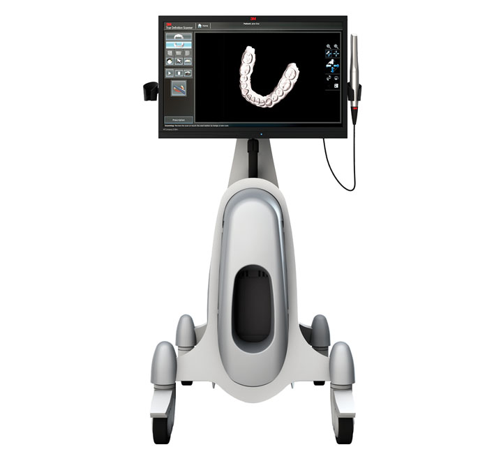 Manufacturers compete to invent increasingly sophisticated [...]
admin2019-09-05T15:07:53+02:00
Innovative solutions in the medical industry
The SMARTTECh4D med scanner is a response to the growing demands of the health and safety sectors. It works with [...]
admin2019-09-05T15:07:48+02:00
Influence of compression socks on swollen legs
In the case of the dimensioning of human physiology, it is sometimes necessary to measure a person and make calculations [...]
admin2019-09-05T15:07:39+02:00
Cosmetic gloves for children and prosthetic funnels
Even 5 to 10 years ago, the prosthetic preparation of the denture funnel, which is the basis of each prosthesis [...]
admin2019-09-05T15:07:30+02:00
The SMARTTECh4D med scanner helps in creating personalized anti-smog masks
The SMARTTECH 3D medical scanner was used in the unique project of creating personalized anti-smog masks initiated by the MASSQ [. ..]
..]
Are you interested? Got questions?
Go to Top
3D scanning in medicine. Types of 3D scanners and areas of use in medicine
Content
-
- Using 3D scanning in medicine
- Advantages 3D scanning
- Types of 3D scanners
- Optical
- Laser
- The main directions in medicine
- Dentistry
- Plastic surgery and cosmetology
- Orthopedics
- Prosthetics
- Student education
Medical use of 3D scanning
3D scanning of a person is necessary for plastic surgeons, dentists, orthopedists and even cosmetologists. Before the advent of 3D technology, doctors had to take measurements manually, as well as create realistic dummies based on the information received: this is a hard and routine job that took a lot of time. 3D scanners have made medical procedures much easier: detailed information about a patient's anatomy can be obtained in minutes.

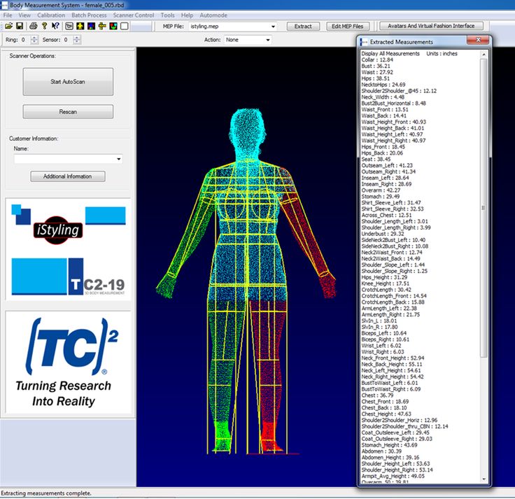
During the scan, the device recognizes the geometry of the body, and the sensors form a cloud of points. Based on the information received, the device software designs a polygonal body model and transfers it to the computer screen. The three-dimensional model becomes the basis for choosing the optimal method of surgical intervention, creating surgical prostheses, orthopedic shoes, and so on.
Benefits of 3D scanning
The 3D human scanner is used in all areas of medicine. Modern 3D scanners are replacing outdated diagnostic methods due to a number of advantages:
-
Three-dimensional models are more accurate and informative. At the diagnostic stage, some medical equipment makes errors, while 3D scanners show the best result;
-
Orthopedic corsets and insoles, created on the basis of a three-dimensional model of the patient, are much more accurate than plaster ones due to the consideration of aspects of individual anatomy;
-
Due to the high speed of work, new technologies allow you to get the necessary information urgently.
 Delay in medicine can cost a person's health and even life, and three-dimensional scanners help prevent the development of undesirable conditions;
 -
Due to its safety, the technology is relevant for the diagnosis of adults and children.
Interesting! 3D scanning is used for cardiovascular surgery. Due to the high accuracy of the information received, surgeons choose the most relevant method of surgical intervention.
Types of 3D scanners
According to the principle of action in medicine, laser and optical 3D scanning is used. Each of these varieties has its own advantages, which we will discuss below.
Optical
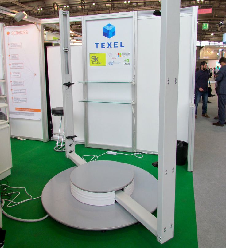
The areas to be examined are illuminated with structured light, while the cameras record the result from different angles. The area is illuminated with a strip of light or a specific pattern (black and white stripes or squares).
 The deformation of the light or pattern conveys to the device information about the shape and depth of the object. The process is recorded by one or two cameras, which transmit information about the structure of the object to the computer software.
 Such devices can be manual and desktop. In the first case, manual control of the device is used, in the second, the scanner is placed on the table. The main advantage of optical devices is the high speed of operation and the ability to transmit color.
Laser
The laser scanner measures the distance to the object under investigation and determines the actual length of the beams. The direction of the laser is controlled by encoders. Based on the analysis of the reflection of rays, a cloud of points is formed, which makes up a three-dimensional image of the object under study. The main advantage of such devices is the ability to digitize objects with a complex shape.
Attention! For the most accurate scan results, the patient must remain static.
Add to compare
Product added to compare Go
Manufacturer Shining 3D Add to compare
Product added to compare Go
Manufacturer Shining 3D Add to compare
Product added to compare Go
Manufacturer Range Vision Add to compare
Product added to compare Go
Manufacturer Range Vision Main directions in medicine
Three-dimensional scanning is actively used in almost all medical fields. The presence of an accurate anatomical model helps to determine the current treatment regimen or to model a prosthesis. We must not forget the importance of virtual models for medical students: such schemes allow us to study the human anatomy in the most detailed way.
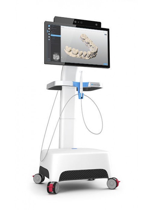
Dentistry
The use of an intraoral 3D scanner for diagnostics
The use of a dental 3D scanner to create a digital model of a plaster jaw prosthesis
Due to oncology, the Australian had to remove 80% of the upper jaw.
 The use of a 3D scanner helped create the perfect jaw prosthesis
 3D dental scanning helps to create detailed virtual pictures of the patient's teeth. Thanks to this, the most complex restoration and restoration work is carried out, orthodontists design prostheses. The technology significantly reduces the time of manufacturing complex structures, and also saves the cost of producing plaster and other models.
The 3D scanner is actively used in dentistry due to a number of advantages. The device is suitable for working with patients with an increased gag reflex. Just a few years ago, to create an impression of the teeth, an impression mass was placed in the patient's mouth: this procedure brought great discomfort. Nowadays, 3D-scanning of teeth is carried out contactlessly and does not bring discomfort.
Attention! The use of a 3D scanner in dental practice has a great advantage: the data obtained during the diagnostics are instantly transmitted to the laboratory.
Plastic surgery and cosmetology
face scan
Familiarization of the patient with the expected result of rhinoplasty based on a three-dimensional image of her face
3D face scanning is actively used in cosmetology and plastic surgery. The study is assigned to study problem areas before carrying out some cosmetic manipulations, as well as to design the expected result of plastic surgery. Thanks to this technology, the patient can get acquainted with how his face will look after surgery. The scanner is used for performing operations on the face (rhinoplasty, blepharoplasty) and body (mammoplasty, abdominoplasty, etc.).
Orthopedics
3D scanning of the spine
3D scanning of the foot to study the anatomical curves
Orthopedic insole printed on the basis of a 3D image of the foot
3D scanning of the body is relevant in the process of determining the type and stage of curvature of the spine or foot.
 The use of x-rays to diagnose the spine and lower extremities does not provide such accurate information, moreover, this procedure is contraindicated in some patients. 3D scanning helps to obtain detailed information about the pathology and develop actual orthopedic products (insoles and corsets).
 For the manufacture of insoles, previously it was necessary to make an impression of the foot. If the doctor made mistakes, the cast turned out to be inaccurate, which affected the quality of the insoles. New technologies help to obtain the most accurate information, regardless of the doctor's actions.
Surgery
3D model of the heart created from scanned data
Siamese twin chest model. Based on the data obtained as a result of the scan, the surgeons managed to perform the most difficult operation to separate them.
The application of new technology plays an important role in surgery. Scanning is aimed at studying the features of the pathology, its location and blood supply.
 This information helps to carry out more accurate and safer operations, to prevent complications during surgery. In addition, scanning is used in the process of bioprinting tissues and organs for transplantation.
 3D scanning is closely related to 3D printing: the surgeon gets the opportunity to study the model of the operated organ or part of the body, develop an actual scheme for surgical intervention and “hone” the movements.
Prosthetics
The use of a 3D laser scanner in transplantology
Prosthesis based on scan data
In the process of creating manual prostheses, it is necessary to accurately determine the location of tissues, muscles and blood vessels. If errors are made during the diagnosis, local necrosis may develop due to tissue rejection. Modern technologies allow collecting the most accurate information about the shape of the stump and the location of tissues to create high-quality and comfortable prostheses.

Student education
A student studies pathological anatomy based on a 3D cadaver model
Scanning helps medical students study anatomy more effectively. The new method is much more efficient than the use of 2D images in classical anatomical atlases. Some programs allow you to observe the surgical intervention: for example, the trajectory of the scalpel during the operation.
Interesting! In France, a 3D scanner is being used to study pathological anatomy. There was no need for an in-person autopsy, and the resulting schemes make it possible to study the human body in layers.
Add to compare
Product added to compare Go
Manufacturer Medit Add to compare
Product added to compare Go
Manufacturer 3Shape Add to compare
Product added to compare Go
Manufacturer ScanPod3D Add to compare
Product added to compare Go
Manufacturer Shining 3D Popular medical 3D scanners
A high-quality three-dimensional scanner provides accurate body diagnostics and successful operations.
 Next, we list a few scanners that have managed to establish themselves in the global market.
Shining Autoscan DS-EX
The device has an open, user-friendly platform that facilitates the scanning process. Various modules are easily installed on the platform, thanks to which the models are scanned in the articulator, the dental impression is examined from various angles, etc. A 3D scanner is used in a dental laboratory to create a virtual patient model, create a treatment plan, design crowns and jaw prostheses. The device is compact and highly accurate.
ScanPod 3D UPOD-S
An orthopedic scanner is used to obtain monochrome and color images. Despite its compactness, the device has a large scanning area, and high speed allows you to use the scanner in emergency mode. In addition, ScanPod 3D UPOD-S is capable of performing 40 operations on the resulting image, and also provides the ability to compare the anatomical features of the right and left foot.
 You can learn more about the features of the scanner in our review.
 Medit i500
The intraoral 3D scanner allows you to scan the entire oral cavity in a matter of minutes. A 3D Full HD color image is displayed on the screen. A small scanning module provides convenience to the doctor during the examination and does not bring discomfort to the patient. Due to its low weight, it is convenient to use even during long-term diagnostics.
3Shape TRIOS 3 Basic Pod
The compact device provides maximum comfort for both doctor and patient during diagnostics. The intraoral scanner works non-contact: no special powders need to be used for the procedure. The resulting color image is sent to the computer screen in high resolution. The CAD/CAM software provides the ultimate in ease of use, and thanks to the USB port, the 3Shape TRIOS 3 Basic Pod can be connected to any computer.
ScanPod3D USOL
ScanPod3D USOL is a compact orthopedic scanner that allows you to quickly and accurately scan the foot of one foot.
 The resulting image can be scaled and rotated for detailed viewing. Diagnostics can be carried out without load, with partial and full load. The device scans the arch of the foot, insoles and lasts, and the RX shape setting is used to create orthopedic shoes.
 Caliber
The portable handheld scanner allows you to get a digital image of objects from 20 cm to 10 m. Weight is 900 g, so operation is not difficult. The touch screen displays the result of the scan, so there is no need for constant checking with the computer. The scanner is used to examine the fine details, and the powerful software ensures easy operation. Calibry is used in 3D scanning of the spine, face, joints and other parts of the body.
Shining 3D EinScan H
The handheld scanner is lightweight and compact, making it easy to carry around. The use of the latest developments in data capture helped to achieve stunning scan results: up to 1,200,000 points per second.
 Shining 3D EinScan H has a number of advantages:
-
Whole body scanning solution;
-
Reliable color reproduction;
- Fast scanning 1200000 points / sec.;
-
High accuracy of 0.05mm scan data.
The new 3D scanner for human scanning provides the most accurate and detailed information about the structure of tissues and organs.
Forecasts of the development of 3D technologies in medical practice
According to International Data Corporation (IDC) forecasts, by 2023 the industry is expected to grow by 635 million units. This means that due to the high accuracy of diagnostic data and the improvement of software, most modern clinics will prefer the use of three-dimensional scanners instead of outdated diagnostic equipment.
Add to compare
Product added to compare Go
Manufacturer Thor3D Add to compare
Product added to compare Go
Manufacturer Shining 3D Add to compare
Product added to compare Go
Manufacturer 3Shape Add to compare
Product added to compare Go
Manufacturer ScanPod3D Expert in the field of additive and subtractive technologies, 3D equipment and CNC machines with over 10 years of experience.

3D scanners in medicine and dentistry
3D scanners in medicine and dentistrySupplier of 3D equipment since 2010
+7 495 646-15-338 800 333-12-82
3D-scanners3D printer Support
On the Project-Propulsion Service
ContactsA 3D scanner is an important attribute of medical research centers and practicing medical institutions around the world. With the help of three-dimensional scanners, it is possible to obtain, for example, an accurate 3D model of the structure of the human body or its individual parts. Plastic surgeons can obtain an accurate color 3D model of the breast, face and any other part of the body in minutes and visually demonstrate the results of future work.
- 3D-innovations in medicine
- Opportunities
- Choosing 3D scanner
- 3D (medicine in medicine
- Possibly
- selection 3D scanner
3D-innovations in medicine
Three-dimensional scanners are successfully used and orthopedists to create high-precision scans of body parts.
 This means that specialists can produce prostheses that are ideally suited to their patients without spending as much money on design as before.
 Previously, the process of producing prostheses and corsets was labor-intensive and uncomfortable. The patient was covered with plaster and waited. After hardening, the gypsum was cut off and sent to production. The manufacturer received the form and manually took measurements.
Now that medical institutions have the opportunity to use 3D scanners, there is no longer a need for expensive and time-consuming work to create plaster models, there is no need to contact the delivery service and wait for the cargo to arrive. Corsets created according to a 3D model are more accurate than plaster corsets, because they take into account all the nuances of the body structure.
Most recently, the design of dentitions took several weeks. Now, thanks to the advent of ultra-precise 3D scanners, the process is simplified and accelerated to several days.

3D innovations in medicine
3D scanners are successfully used by prosthodontists and orthopedists to create high-precision scans of body parts. This means that specialists can produce prostheses that are ideally suited to their patients without spending as much money on design as before.
Previously, the process of producing prostheses and corsets was labor-intensive and uncomfortable. The patient was covered with plaster and waited. After hardening, the gypsum was cut off and sent to production. The manufacturer received the form and manually took measurements.
Now that medical institutions have the opportunity to use 3D scanners, there is no longer a need for expensive and time-consuming work to create plaster models, there is no need to contact the delivery service and wait for the cargo to arrive. Corsets created according to a 3D model are more accurate than plaster corsets, because they take into account all the nuances of the body structure.
Most recently, the design of dentitions took several weeks. Now, thanks to the advent of ultra-precise 3D scanners, the process is simplified and accelerated to several days.
Capabilities
Placement of prostheses
- Scan any part of the body to create functional prostheses
- 3D archiving - no need to store multiple castings
- Modeling and planning in plastic surgery
- Orthopedics and cosmetology, dentistry
Diagnosis of the development of the skull of children
- Scanning of the head of a child to fit a soft helmet
- Subsequent head scan to track change
Wound care
- Color and texture wound scanning for quick selection of treatment methods.
-