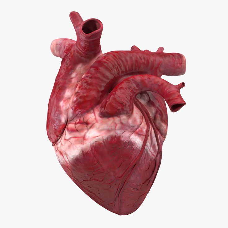
3D print anatomical heart
Collection: Heart Library | NIH 3D Print Exchange
Image Credit: Dr. Matthew Bramlet; Jump Trading Simulation and Education Center.
Welcome to the 3D Heart Library, a collection of digital reproductions of human anatomic hearts, with a specific area of interest in congenital heart disease. The highly accurate 3D representations of hearts have been created directly from patient MRI data. The goal of the 3D Heart Library is to serve as a platform for sharing, collaboration, and education.
If you are a medical or engineering professional with questions about the 3D Heart Library or are interested in submitting a case study, please complete the contact form on the Jump Simulation website.
More models and info are on the way as this exciting new collaboration develops!
Search
Diagnosis/Condition
- Any -Normal heartSeptal Defects-Atrial Septal Defect (ASD)--PFO--Secundum--Primum--Sinus venosus--Coronary sinus--Common atrium (single atrium)-Ventral Septal Defect (VSD)--Type 1 (Subarterial) (Supracristal) (Conal septal defect) (Infundibular)--Type 2 (Perimembranous) (Paramembranous) (Conoventricular)--Type 3 (Inlet) (AV canal type)--Type 4 (Muscular)--Gerbode type (LV-RA communication)--Multiple-AV Canal--AVC (AVSD) Complete (CAVSD)--AVC (AVSD) Intermediate (transitional)--AVC (AVSD) Partial (incomplete) (PAVSD) (ASD primum)-AP Window--Aortopulmonary window--Pulmonary artery origin from ascending aorta (hemitruncus)-Truncus Arteriosus--Truncus arteriosus + Interrupted aortic arch--Truncal valve insufficiencyPulmonary Venous Anomalies-Partial Anomalous Pulmonary Venous Connection--Scimitar-Total Anomalous Pulmonary Venous Connection--Type 1 (supracardiac)--Type 2 (cardiac)--Type 3 (infracardiac)--Type 4 (mixed)Cor TriatriatumPulmonary Venous StenosisSystemic Venous Anomalies-Anomalous Systemic Venous Connection--Systemic venous anomaly-Systemic venous obstructionRight Heart Lesions-Tetralogy of Fallot--Pulmonary stenosis--AVC (AVSD)--Absent pulmonary valve-Pulmonary Atresia--IVS--VSD (Including TOF PA)--VSD-MAPCA (pseudotruncus)--MAPCA(s) (major aortopulmonary collateral[s]) (without PA-VSD)-Tricuspid Valve Disease and Ebsteins Anomaly--Ebstein's anomaly--Tricuspid regurgitation non-Ebstein's related--Tricuspid stenosis--Tricuspid regurgitation and tricuspid stenosis--Tricuspid valve - Other-RVOT Obstruction and/or Pulmonary Stenosis--Pulmonary stenosis - Valvar--Pulmonary artery stenosis (hypoplasia) - Main (trunk)--Pulmonary artery stenosis - Branch - Central (within the hilar bifurcation)--Pulmonary artery stenosis - Branch - Peripheral (at or beyond the hilar bifurcation)--Pulmonary artery - Discontinuous--Pulmonary stenosis - Subvalvar--DCRV-Pulmonary Valve Disease--Pulmonary valve - Other--Pulmonary insufficiency--Pulmonary insufficiency and pulmonary stenosisShunt failureConduit failureLeft Heart Lesions-Aortic Valve Disease--Aortic stenosis - Subvalvar--Aortic stenosis - Valvar--Aortic stenosis - Supravalvar--Aortic valve atresia--Aortic insufficiency--Aortic insufficiency and aortic stenosis--Other-Sinus of Valsalva Fistula/Aneurysm--Sinus of Valsalva aneurysm-LV to Aorta Tunnel-Mitral Valve Disease--Mitral stenosis - Supravalvar mitral ring--Mitral stenosis - Valvar--Mitral stenosis - Subvalvar--Mitral stenosis - Subvalvar, Parachute--Mitral stenosis--Mitral regurgitation and mitral stenosis--Mitral regurgitation--Mitral valve - Other-Hypoplastic Left Heart Syndrome (HLHS)-Shones syndromeCardiomyopathy-Cardiomyopathy (including dilated, restrictive, and hypertrophic)-End-stage congenital heart diseasePericardial Disease-Pericardial effusion-Pericarditis-OtherSingle Ventricle-DILV-DIRV-Mitral atresia-Tricuspid atresia-Unbalanced AV canal-Heterotaxia syndrome-Other-Single Ventricle + Total anomalous pulmonary venous connection (TAPVC)Transposition of the Great Arteries-Congenitally Corrected TGA-Congenitally corrected TGA + IVS-Congenitally corrected TGA + IVS-LVOTO-Congenitally corrected TGA + VSD-Congenitally corrected TGA + VSD-LVOTO-IVS-IVS-LVOTO-VSD-VSD-LVOTODORV-VSD type-TOF type-TGA type-Remote VSD (uncommitted VSD)-DORV + AVSD (AV Canal)-DORV, IVSDOLVThoracic Arteries and Veins-Coarctation of Aorta and Aortic arch hypoplasia--Coarctation of aorta--Aortic arch hypoplasia--VSD + Aortic arch hypoplasia--VSD + Coarctation of aorta-Coronary Artery Anomalies--Anomalous aortic origin of coronary artery from aorta (AAOCA)--Anomalous pulmonary origin (includes ALCAPA)--Fistula--Aneurysm--Other-Interrupted Arch--Interrupted aortic arch--Interrupted aortic arch + VSD--Interrupted aortic arch + AP window (aortopulmonary window)-Patent Ductus Arteriosus--Patent ductus arteriosus-Vascular rings and Slings--Right arch aberrant left subclavian artery--Double aortic arch--Circumflex aortic arch--Pulmonary artery sling-Aortic Aneurysm--Aortic aneurysm (including pseudoaneurysm)-Aortic DissectionMiscellaneous-Aneurysm--Ventricular Right (including pseudoaneurysm)--Ventricular Left (including pseudoaneurysm)--Pulmonary artery--Other-Dextrocardia-Cardiac tumor-Endocarditis-Hypoplastic LV-Hypoplastic RV-Cardiac - Other-Atrial Isomerism--Left--Right-Levocardia-Pulmonary vascular obstructive disease (Eisenmengers)-Pulmonary AV fistula-Rheumatic heart disease-Situs inversus-Mesocardia-Postoperative bleeding-Prosthetic valve failure-Myocardial infarction-Primary pulmonary hypertension-Persistent fetal circulation-Status post - Transplant--Heart--Lung(s)--Heart and lung-Other
Segmentation Method
- Any --Myocardium-Solid blood pool-Blood pool border
Affiliation
'Augusta UniversityBarry University School of Podiatric MedicineBINZHOU MEDICAL UNIVERSITY HOSPITALBoston University School of MedicineBrigham and Women's HospitalBrooklyn Department of Emergency Medicine has been working with me for the design and prototyping processCase Western Reserve University School of MedicineChildren's Hospital of IllinoisChildren's National HospitalDecatur Memorial HospitalDepartment of Veteran Affairs - VA BronxDivision of Surgery and Interventional Sciencee-NABLEEmbodi3DHarvard Medical SchoolHospital Quiron VallèsIlumens Paris DiderotINDIANA UNIVERSITYInstituto Nacional de Salud del Niño. LimaIsrael & Stony Brook UniversityKennebec Valley Community Collegekennedy kreigerKing's County Hospital/SUNY DownstateLearning Resource CenterMassachusetts General HospitalMaterialiseMayo Clinic RochesterMcGuire Veteran's HospitalMedical College of GeorgiaMeSegmenter.comMidwestern UniversityMySegmenter.comN/ANational Institutes of HealthNIH Clinical CenterNoneNova Southeastern University Health Professions Division LibraryNRICPNYOhio Universityonmouseover=OSFPanorama Centre for Surgical OncologyPenn VetPeruPhilipps University of MarburgPOLIRADIO23Pontificia Universidad Católica de ChilePreston Smith LibraryRabin Medical CenterRochester General HospitalRowan UniversityRoyal Free HospitalSheba Medical CenterSibley Memorial HospitalSingaporeStellenbosch UniversityTest HospitalTexas Tech Health Science CenterThe Cancer Imaging ArchiveThomas Jefferson UniversityTTUHSCTTUHSC // UMCUCSF Benioff Children's HospitalULTRAMEDUniformed Services University of the Health SciencesUniversity at BuffaloUniversity College LondonUniversity of AlbertaUniversity of California San FranciscoUniversity of Colorado School of MedicineUniversity of MassachusettsUniversity of Rochester Medical CenterUniversity of Test StateUniversity of Toronto/Toronto General HospitalUniversity of VirginiaUniversity of WashingtonUniversity of Washington Medical CenterUS Food and Drug AdministrationUSAVA Sierra Nevada Health Care SystemVeterans Innovation Challenge
Materialise HeartPrint | 3D-Printed Cardiovascular Models
3D-printed cardiovascular models
Gain better insight into complex pathologies with realistic, 3D-printed heart models.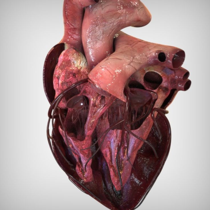 Get a deeper understanding of complex spatial relationships between different structures by using an exact replica of your patient’s anatomy. Transform your patient’s experience and increase their understanding by using the latest 3D planning and 3D printing technology.
On this page:
Communicate more clearly
Discuss heart conditions more precisely with your multidisciplinary team, patients, and their families to achieve true informed consent.
Educate with detailed models
Use the latest 3D printing technology to educate and train the whole heart team on complex anatomy, new procedures, and the latest device innovations.
Precise anatomical representation
Get a one-to-one replica of your patient’s anatomy and appreciate the spatial understanding in 3D thanks to accurate segmentation tools.
Improve patient selection in clinical trials
Perform tests with personalized and anatomically accurate models and increase the chance of success in clinical trials by improving patient selection and planning.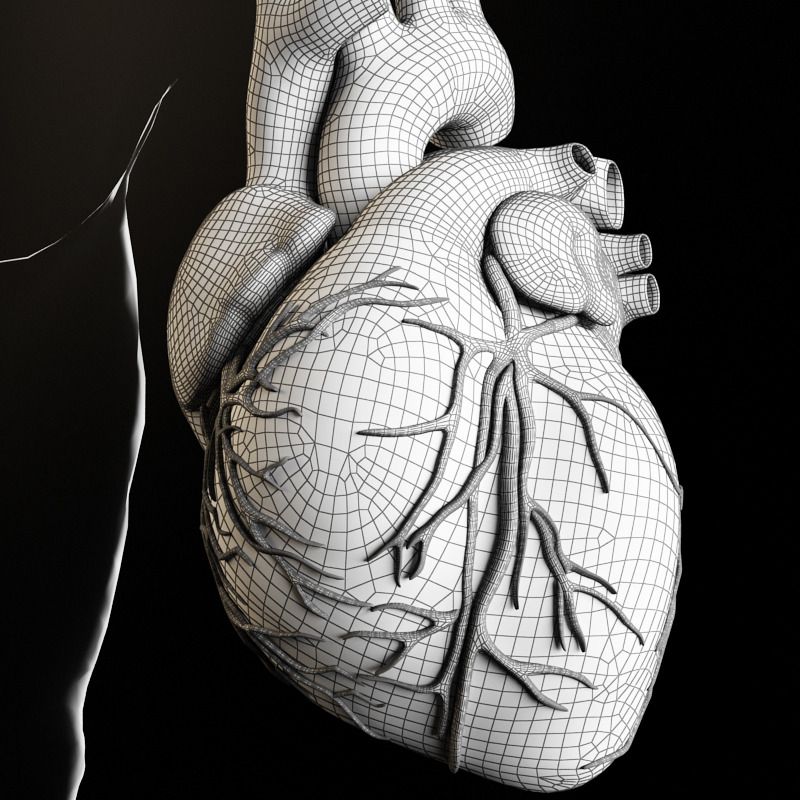
Communicate more clearly
Communicate clearly with physicians to ensure they’re better equipped to prepare for cutting-edge procedures.
Avoid surprises in the cath lab
Ensure that physicians are fully prepared before entering the cath lab to avoid surprises and minimize on-the-spot decision-making.
Material and color options
HeartPrint Flex
Premium material to best represent the compliance of cardiac tissue. Useful for creating flexible representations of anatomy to create a more realistic planning and training experience.
Discover where HeartPrint makes an impact
Cardiovascular medical devices
Structural heart
“I think 3D-printed models offer a completely new method for testing our instruments, allowing them to be tested in scientifically valid models that can be easily extended with additional requirements.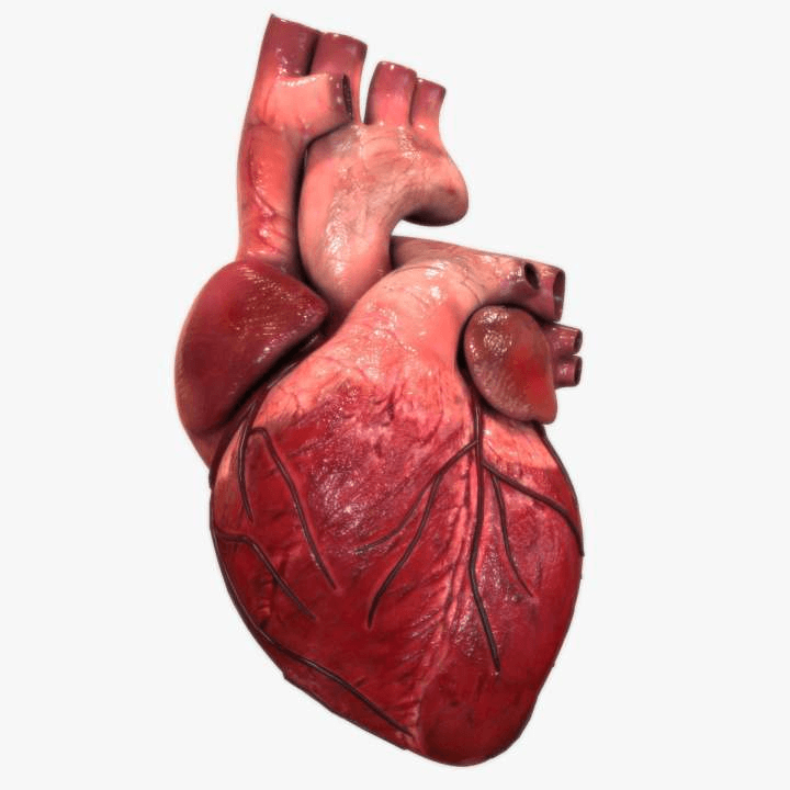 ”
”— Ir. Awaz Ali, Bio-Inspired Technology research group, TU Delft
Interested in Materialise HeartPrint?
Do you have any questions, or are you interested in learning more about Materialise HeartPrint? Get in touch with us, and let’s discuss how we can support your work.
Talk to an expert
Medical sales team
Locate your nearest officeEmail [email protected]Learn more and get support
Find out how to get started with HeartPrint in our academy and get customer support with the following links.
Quick links
Explore our Academy resourcesView material characteristicsImaging guidelinesWHITEPAPER
Material Characterization of Materialise Heartprint Models and Comparison
CASE STUDY
3D-Printed Heart Model Helps 16-Year-Old Heart Tumor Patient
2 min read
WHITEPAPER
Material Characterization of Materialise Heartprint Models and Comparison
CASE STUDY
3D-Printed Heart Model Helps 16-Year-Old Heart Tumor Patient
2 min read
Read all regulatory information
L-102743-01
Materialise medical device software may not be available in all markets because product availability is subject to the regulatory and/or medical practices in individual markets.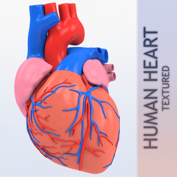 In countries where no regulatory registration is obtained of Mimics and/or 3-matic Medical, a research version is available. Please contact your Materialise representative if you have questions about the availability of Materialise medical device software in your area.
You might also like
Mimics Enlight TMVR
Expert structural heart planning with true 3D clarity for transcatheter mitral valve replacement procedures
Mimics Enlight LAAO
Expert structural heart planning with true 3D clarity for left atrial appendage occlusion procedures
Materialise Mimics inPrint
From scan to 3D model with ease
Mimics Innovation Suite
Industry-standard software for segmentation and anatomical analysis, planning, and design
© Copyright Materialise 2023
Cookie Statement
Legal terms
Privacy notice
Living heart printed on a 3D printer - Kommersant FM - Kommersant
In Israel, for the first time in the world, a living heart was created on a 3D printer, which consists of tissues and blood vessels, and also has cameras.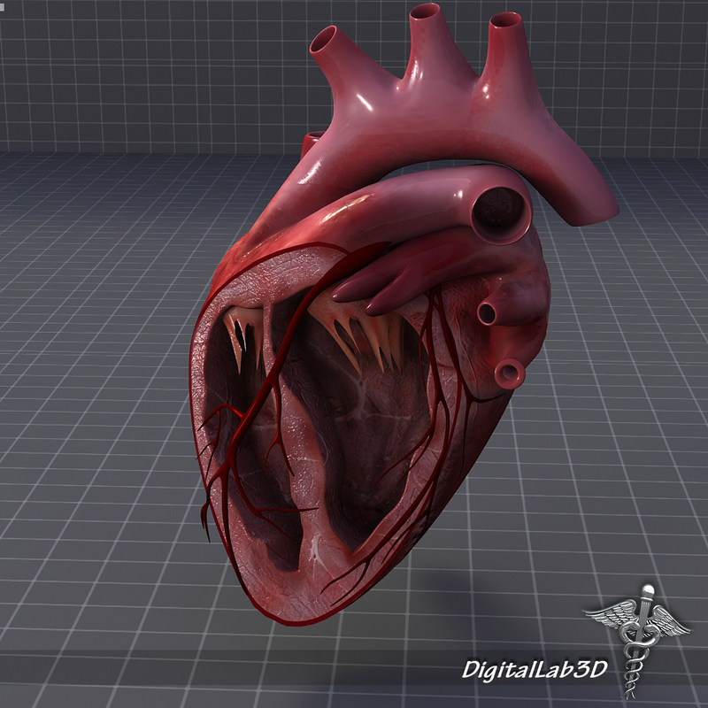 True, at the moment it can only suit a rabbit because of its small size. But scientists from the University of Tel Aviv are confident that in the future they will be able to print a heart for a person. How did you manage to create an organ on the printer? And can we talk about a revolution in medicine? Anna Nikitina and Gleb Silko will tell.
Photo: Yaroslav Chingaev, Kommersant / buy photo
The world's first 3D-printed heart resembles a berry: its size is about 2.5 cm, although it took more than three hours to print. However, even now the achievement of Israeli scientists is called a medical breakthrough. The heart is made from human fat cells and connective tissue. Previously, synthetic substances were used for this.
In the future, this new technology will not only solve the problem of the shortage of organs for transplantation, but will also facilitate the transplantation process as much as possible, says Israeli journalist Sasha Vilensky: “The most global problem is the rejection of a transplanted organ by the body.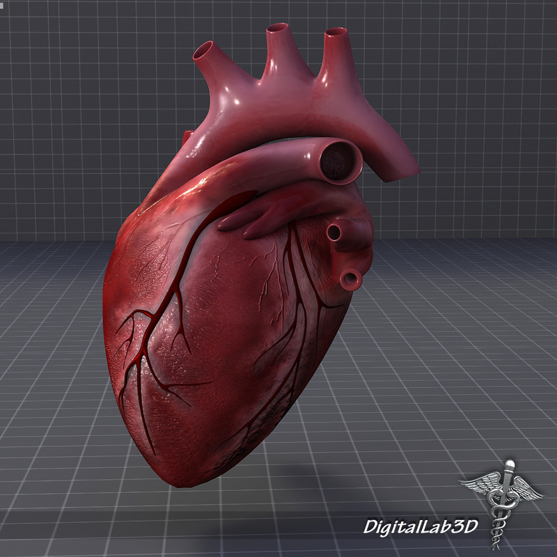 There is always such a danger. But the whole point of interest is that in the case of a 3D printer, we are dealing with what is grown and printed from the cells of the patient himself, thus simply removing the issue of rejection. This is genius".
The problem of organ shortages around the world is really acute. In Russia, for example, in 2017, there were only 900 donors per 6 million people. And the most complex organ - the heart - was transplanted 250 times in a year, while it required almost 2 million people. The experience of Israeli scientists in printing hearts is impressive, and it can be used in other countries, says Yousef Hesuani, executive director of the 3D Bioprinting Solutions laboratory. True, according to him, it is still too early to talk about a revolution in medicine: “Scientists have used a very interesting material based on collagen - this is a protein in the body of mammals. In addition, it is from the point of view of creating a complex three-dimensional structure that the researchers are great, they did a really good job. However, the form does not yet determine the function, especially if we are talking about such a complex organ as the heart.
New approaches have been used, but unfortunately it is too early to say that there has been an incredible breakthrough in this area.
When, for example, the native organ is removed and a new one is transplanted, and the function is fully restored, this will become an unconditional revolution.”
Tel Aviv University itself, where the heart was printed, says that in the future the necessary organs can be printed directly in hospitals. Now similar developments are being carried out all over the world. Russian companies, for example, are trying to create an artificial liver and kidneys. True, this process is too expensive, and it is unlikely that in the near future the technology will be introduced on a mass level, says the director of the National Medical Center for Transplantation and Artificial Organs. Shumakova Sergei Gauthier: “What the Israeli doctors did is just great. This proves that these possibilities can really be used to create some kind of anatomical structures. As for other organs, such as the kidney and liver, our institute is also working on this. We also have bioprinters, but frankly, growing such tissue complexes is a rather complicated and expensive technology, which is very different from the traditional method of organ transplantation.”
There are already examples in the world when doctors managed not only to create artificial organs on a 3D printer, but also successfully transplant them, for example, a few years ago in Russia a thyroid gland was printed, which took root in a patient. And last year, scientists from Jerusalem implanted a prosthesis for the skull on a patient, and it was also created using three-dimensional technologies.
During this year, Israeli scientists plan to test printed hearts on rabbits and rats, and then start working on creating a human heart.
Dipol Press Center
April 23, 2019
subscribe subscribe
Printing a heart on a 3D printer - a few years ago, only science fiction writers could afford to use such a phrase.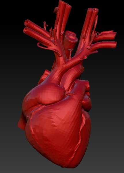 However, scientists from Tel Aviv University (Israel) managed to make a real technological breakthrough: for the first time in the world, they created a human heart using a medical 3D printer.
The adipose tissues of one of the patients served as the basis for creating the main organ of the human body artificially. Scientists have reprogrammed cell structures so that they turn into artificial stem cells, from which various types of tissues are formed in the human body. And connective tissues - glycoproteins - were processed into "ink" for a medical 3D printer in the form of a personalized hydrogel.
This is the first time this technology has been used: previous experiments to create an artificial heart using a printer involved the use of synthetic materials. The same organ has all the immunological, cellular, biochemical and anatomical properties characteristic of the human body.
The size of the printed heart is no larger than that of a rabbit. But it has everything necessary for life - blood vessels, ventricles and chambers. The cells of the new heart even contract, but not yet synchronously with each other, so the artificial organ is not able to pump blood, this has yet to be achieved.
According to Israeli scientists, an artificial human heart of normal size, which will begin to beat for real and perform all its functions, will be created using this technology not earlier than in 5-10 years. However, in the next year or two, they plan to start experiments on animals, transplanting them with hearts printed on a printer.
End-stage cardiovascular diseases provide only one way to save the patient - heart transplantation. However, there is an acute shortage of donors for such complex operations, and patients have to wait a long time for a transplant. Given these circumstances, 3D printing of the heart opens up new perspectives in the field of transplantation, which will save the lives of many patients.