Fit 3d body scanner
Know Your Body Composition & Body Fat
To help provide the absolute best member experience, Spunk Fitness has partnered with FIT-3D, the world’s leader in 3D body scanning technology.
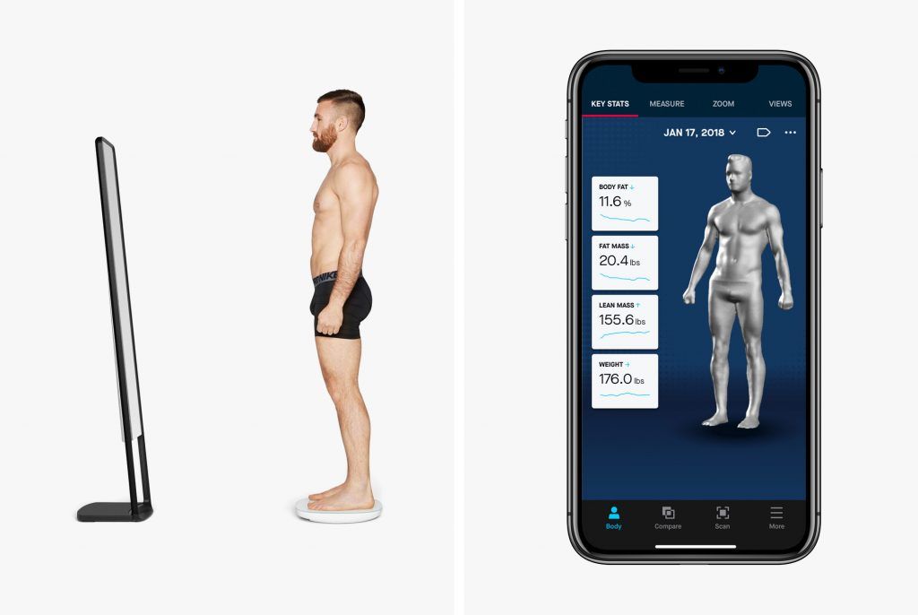
In a single 40 second, non-invasive scan, members can capture circumference measurements, body composition, posture analysis, Body Shape Rating (BSR), Basal Metabolic Rate (BMR) and much more. It also shows you how you compare against healthy ranges and against others of your same gender and age.
Plus, a 3D avatar to help you visually track your progress from any device anywhere in the world. You will be able to compare your baseline scan to any future scan so you can easily monitor, track and see the progress you’re making toward your fitness goals!
As a Spunk Fitness member, you will get a FIT-3D full body scan, a $200 value – at no additional cost to you with each fitness consultation purchased.
See how the FIT-3D Body Scan woks by clicking HERE txhd.io/s/#49iV
Sign-up for your FIT-3D Body Scan HERE txhd.io/s/#QbJA
Fit3D Posture Analysis Identifies Problems That Impact Mobility
I wish I had a dime for every time my Dad said to sit up straight at the table, or my Mom told me to keep my shoulders back. They wanted me to have good table manners and look confident, but perhaps they also knew what science has now taught us: good posture has many health benefits and poor posture can severely limit your mobility — particularly as you age.
Good posture keeps your bones and joints in good alignment and reduces wear and tear on joint surfaces. It also decreases the stress on ligaments. According to Livestrong.com, good posture improves the efficiency of oxygen flow to your nervous system, organs, and other tissues. It can help prevent future health issues like spinal disk problems and constricted blood vessels and nerves. Muscle & Fitness also points out that good posture improves digestion and reduces acid reflux because it enables organs to sit in their natural position without compression.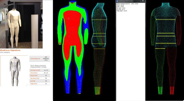
How is Your Posture?
Most people think little about their posture until they begin having chronic back or neck pain. A posture analysis can help identify your risk for problems like kyphosis (commonly known as roundback) that affect mobility. According to the University Spine Center, sufferers of Kyphosis often slouch and struggle with breathing problems, back pain and tenderness, and stiffness. The effects of which can be reduced with physical therapy and exercise.
You may be unwittingly standing, walking, or sitting in ways that will have a significant impact on your mobility. Let’s begin with your hips. The Fit3D Posture Analysis will identify if you have, for example, an anterior pelvic tilt. This is most common in people whose femurs are rotated, walk knock-kneed, and/or have flat feet. Anterior pelvic tilt causes hip, knee, and lower back pain. It will also cause a greater curvature of the spine (kyphosis), which will put you at risk for shoulder and neck pain, and headaches.
Your knees are also vulnerable to the effects of poor posture. A posture analysis can help you identify health problems like over-pronation of the feet, which can cause your knees to fall inwards. According to Ask the Trainer, this is a common sports injury caused by poor posture. The Foot Posture Center warns that the “majority of knee pain cases are directly related to poor foot posture and it is vital that this is corrected naturally before opting for medication or surgery.”
Finally, and perhaps most common, poor posture causes shoulder problems. The shoulder is made up of three bones, the clavicle (collarbone), scapula (shoulder blade), and the humerus (arm bone) that are held together by the rotator cuff. As any of you who have had shoulder injuries know, the shoulder is a complex and delicate joint. Poor posture frequently causes shoulder impingement — which not only causes pain — it can make it difficult for you to raise your arms above your head. If unidentified and untreated, it will wear down the tendons in the shoulder (called bursa).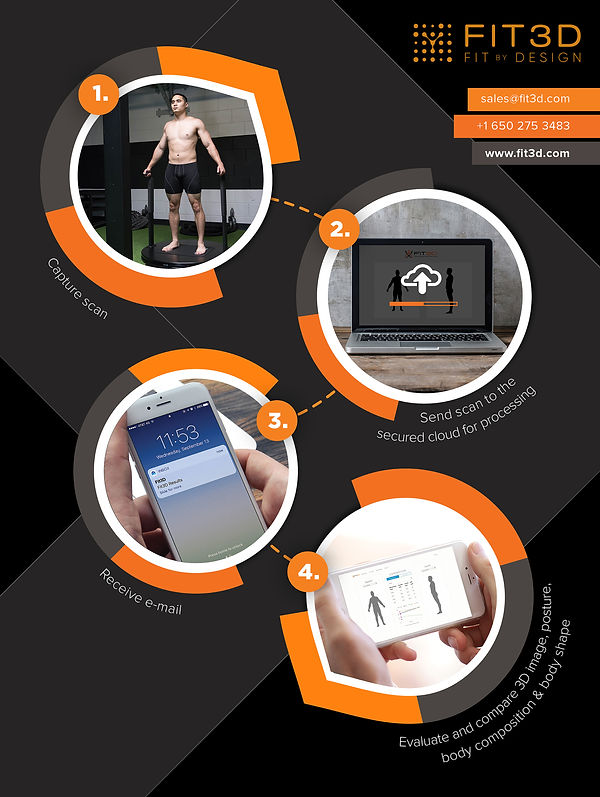 A Fit3D Posture Analysis can get you ahead of all these risks.
Fit3D Posture Analysis
Why Fit3D for Your Posture Analysis?
As part of the 3D body surface scan, the Fit3D ProScanner includes an assessment of your full body posture, from eyes and ears to feet. Unlike other tools that evaluate posture, the Fit3D ProScanner analyzes the position of your ears and eyes as a baseline, which provides important insight into your head and neck positioning. Our Posture Analysis evaluates your shoulders, hips, knees, and ankle positions when you are standing. It also provides posture Wellness Metrics on body shape, waist circumference, waist to hip and trunk to leg volume ratios, and more.
This is particularly important for people with mobility issues, chronic neck, back, and other joint pain. A posture analysis can help you focus your time in the gym and during physical therapy on the right activities to strengthen your core. Over time, as your fitness and health improve, you can take repeat scans to see changes and further calibrate your posture.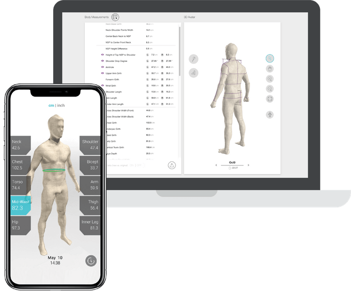 The Fit3D ProScanner Posture Analysis is the only one of its kind that visualizes your changes with an avatar.
Lastly, and perhaps most importantly for your staff, the Fit3D ProScanner automates the entire process of analysis. Using the app with an intuitive interface, our system provides a report with an in-depth level of analysis that would typically require a clinician to determine.
Body Shape Rating (BSR) is a wellness score based on the relationship of your body shape to cardiovascular-related risk factors. It answers the question, is my body shape making it more likely for me to potentially develop certain cardiovascular related health issues? BSR is on a 0-100 range where 50 is average and the higher the score the lower the risk.
Trunk to Leg Volume Ratio compares the volume of your trunk with the volume of your legs. Research claims that having a high percentage of your body’s volume in your torso compared to your legs increases the likelihood of you experiencing prediabetes, diabetes, high triglyceride (fat) counts, high blood pressure, metabolic syndromes, and other severe health complications.
Weight in Your Midsection is highly correlated with visceral fat, which is the unhealthy fat around your organs. This can lead to many negative health effects. If you have a big belly and smaller legs, it is a good assumption that your weight is centered around your midsection and is, therefore, visceral fat.
Ways to Improve
The BSR formula is dependent on volume, not weight. The primary way to improve your BSR is to increase the density of your body by increasing the muscle in your legs and decreasing your waist circumference. This can be done through a balanced mix of good nutrition and exercise. Consult with your trainer, nutritionist, coaches, or doctors to set up a plan.
Body Fat Percentage (BFP) is the user’s total fat mass divided by the total body mass. Fat is an essential component of the body and is necessary to maintain life and reproductive functions; however, too much body fat can negatively affect overall health or hormone levels. It also serves as an indicator of more serious health problems that could potentially be faced in the future.
Fat Mass
- Fat mass is also known as adipose tissue within the body. This loose connective tissue is primarily composed of adipocytes (fat cells). Its main role is to store energy in the form of lipids. It also cushions and insulates the body.
- Body fat is primarily made up of three types of fat:
-
- Epicardial Adipose Tissue (EAT): EAT is a particular form of visceral fat deposited around the heart and is found to be a metabolically active organ that generates various bioactive molecules, which may significantly affect cardiac function.
- Subcutaneous Fat: The fat that is generally stored just below the surface of the skin. Subcutaneous fat is not related to many of the classic obesity-related pathologies, such as heart disease, cancer, and stroke and is generally a protective fat.
- Ectopic Fat: The storage of triglycerides (fats) in tissues other than adipose tissue that are only supposed to contain small amounts of fat, such as the liver, skeletal muscle, heart, and pancreas.
 These fats can interfere with cellular function. The cause for accumulation of Ectopic fat is unknown.
Ways to Decrease Body Fat
You can decrease your body fat percentage through a balanced mix of good nutrition and exercise that includes higher intensity interval training and cardio. Consult with your trainer, nutritionist, coaches, and/or doctors to set up a plan.
Lean Mass is the muscle tissue, skeletal tissue, and water in the body
Ways to Increase Lean Mass
You can increase your lean mass by participating in health and fitness programs that include weight training. You can also talk with your nutritionist to develop a nutrition program that will effectively build lean mass. If you are simply looking to build lean mass, but not necessarily lose fat mass, your program may be quite different than a program that is geared towards a more balanced fat percentage reduction. Consult with your trainer, nutritionist, coaches, and/or doctors to set up a plan.
Basal Metabolic Rate (BMR) is the amount of energy expended while at rest in a neutrally temperate environment and in the postabsorptive state. In this state, the digestive system is inactive, which is usually achieved by about twelve hours of fasting. Furthermore, BMR is the amount of energy expressed in calories that a person needs to keep the body functioning (i.e. breathing, blood circulation, controlling body temperature, cell growth, brain and nervous function, and contraction of muscles). BMR affects the rate at which a person burns calories and ultimately affects whether that individual maintains, gains, or loses weight.
Ways to Increase BMR
You can increase your BMR by participating in fitness programs that include high-intensity interval training or lifting weights to grow muscle. You can further increase your BMR by eating more protein, eating smaller-portioned meals per day, and staying hydrated. The rule of thumb here is that muscle requires more energy to survive—the more lean muscle you have on your body, the higher your BMR. Consult with your trainer, nutritionist, coaches, and/or doctors to set up a plan.
Fit 3D Body Scanning | G&M Medical Center
We all struggle with body issues from time to time, whether clothes aren’t fitting as well as they used to or the body has gone through changes in recent months. Fortunately, the digital age brings us one of the greatest inventions yet: Fit 3D body scanning. This cutting-edge technology produces 3D replicas of your body so you can track changes over time, and it is available at our luxury medspa.
What is Fit3D ?
The entire process begins with stepping onto our 3D body scanning platform, grabbing onto the handles, and standing as still as you can for about 40 seconds. The platform will rotate as the sensor moves up and down, scanning every inch of your body to get the most accurate results. In less than a minute, the Fit3D Proscanner will extract more than 400 measurements including circumferences, heights, lengths, widths, volumes, and surface areas from your entire body.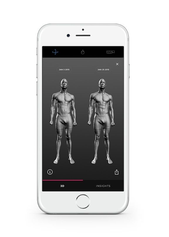 The most important of these measurements are mapped onto a 3D image and can be used in wellness assessments to show progress and analyze what needs improvement. A comprehensive report is emailed to you within 5 to 10 minutes of scan completion, and our trained technicians are always available to answer questions regarding the scan and information gathered as well.
What is Fit 3D Body Scanning Used For?
As 3D body scanning becomes more mainstream, the uses for this technology are increasing by leaps and bounds, as well. Fit 3D body scanning is probably best known for its applications in the health and fitness industry. Wellness and fitness centers use Fit 3D body scanning technology to accurately depict the client’s measurements and highlight areas of improvement. During the fitness regimen, Fit 3D body scans can track changes in BMI and body shape by comparing recent body scans to older body scans. This process can continue as long as it takes for the client to reach his or her goal, whether through personal training for fitness or weight loss programs.
Similarly, aesthetic practices use Fit 3D body scanning to verify the efficiency of their body contouring programs. Before a client starts a body contouring journey, a certified aesthetic technician creates a 3D model of the client’s body. Once the program is finished, another replica is created to compare the before-and-after results.
Furthermore, Fit 3D body scanning is being extended into the fashion industry. Ordering custom fitting garments can be a frustrating process, especially if you don’t have access to a tailor. With an accurate digital depiction of your measurements, you can easily have clothes custom-made to fit your body shape perfectly. The Fit 3D program will automatically send your measurements to the StitchFix app to create your perfect fit. All you have to do is sit back and enjoy your new look.
If you’re looking to learn more about Fit 3D body scanning, please don’t hesitate to give us a call! A
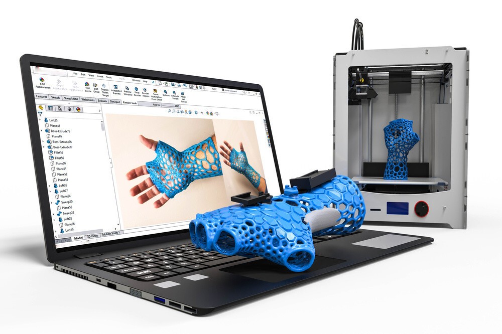
how to quickly create prostheses and other unique products
Reverse engineering
Implementation stories
Medicine
Author: Aleksey Chekhovich
Author: Aleksey Chekhovich
Video: Real-time 3D Face Scan | Order a free test scan | 3D scanner helps victims | 3D scanning of the human body in great detail
Creating prostheses using 3D technologies: the era of new possibilities
As technology improves and becomes more accessible, there are new opportunities to solve the most complex medical problems.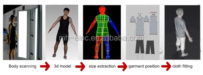 A team of researchers from the company Chabloz Orthopedie (France) managed to create a unique and truly revolutionary prosthesis.
Chabloz Orthopédie worked with Denis Gauthier, who had his forearm amputated. First, the experts performed a 3D scan of the patient's healthy arm using a peel 3d scanner to obtain its mirror image. This was done so that the designed product ideally repeated the proportions of a healthy limb. The rest of the amputated arm was also scanned to achieve a comfortable and ergonomic fit for the prosthesis.
Denis Gaultier with finished prosthesis
The team then proceeded to design a CAD model and develop a myoelectric prosthesis. Batteries, sensor cables and an artificial hand were integrated with the fabricated forearm. The prosthesis itself was designed in CAD and printed on a 3D printer. HP Jet Fusion technology was used to print various components of the forearm. After manufacturing, all parts were finished and painted.
The use of 3D scanning and 3D printing guarantees not only the correct fit of the fixture, but also gives complete freedom of movement.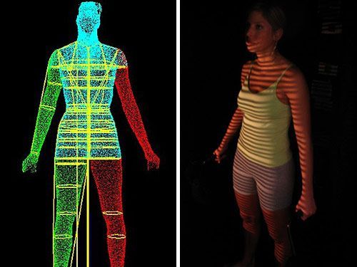 A new approach to prosthesis design allows professionals to develop solutions that are lightweight and highly customizable. Did you know that 3D printed parts are 20% lighter than their carbon or fiberglass counterparts? Such products also have the necessary rigidity, hardness and durability.
Later, this innovative prosthesis was combined with the BeBionic hand, one of the most advanced bionic limbs, and a state-of-the-art myoelectric forearm and hand was custom-made. Gauthier's case is a great example of the innovative use of 3D measurement and 3D printing technologies.
The video shows the complete process of digitizing the face and ears using the Creaform Go!SCAN 20 portable 3D scanner (its analogue in white peel 2 is now being produced). This solution provides a detailed digital model for use in areas such as plastic and reconstructive surgery, in particular, facial prosthetics.
Order a 3D test scan for free!
3D scanner helps victims
Unfortunately, tragedies inevitably occur around the world, and those affected need help. But, fortunately, there are organizations such as Médecins Sans Frontières (Doctors Without Borders) that do their best to provide them with the necessary treatment and care. Since 2016, the organization has been working tirelessly to solve the problem of providing prostheses to those in need around the world. The goal is to help amputees regain independence. The solutions that are used in this case are 3D technologies.
Upon completion of the medical examination of the patient, doctors determine his needs and expectations. Using the peel 3d scanner, with minimal discomfort for the victim, a high-precision digital model of the injured limb is created.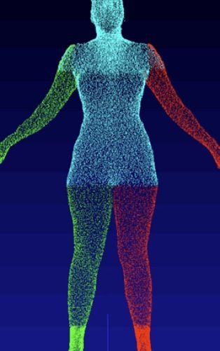 Compared to traditional impression making, 3D scanning is significantly faster and does not require contact. The result of the 3D scan is then transferred to the virtual sleeve and prosthesis design software. The developed component is made on a 3D printer and installed on the patient's limb. If necessary, during the installation process, the prosthesis and sleeve are finalized. After three months of use, the patient is invited to evaluate the comfort of the prosthesis.
MSF also developed a similar procedure to create compression masks for burn patients. Here, the use of non-contact technology also provides significant advantages over traditional plaster bandages, not only making the procedure less painful for the patient, but also speeding it up, allowing for a much larger number of people in needy countries to be treated.
Physiotherapist Pierre Moreau scans a patient with head burns. Then, based on the scan, a transparent pressure therapy mask is created / Photo: Elisa Oddone, Al Jazeera
Pierre Moreau, a physiotherapist from Médecins Sans Frontières, says: “The purpose of the 3D project is to help patients in need of special rehabilitation care. We launched this project in Amman in 2017, where we started providing upper limb amputees with 3D printed prostheses. But simply printing a prosthesis is not enough - we try to understand the needs of the victims, find individual solutions and keep them in their new position for as long as possible. We are assisted by an interdisciplinary team of rehabilitation workers and engineers. Then our specialists helped more than 30 patients in Jordan.
In 2018, we began to use the technology in another direction - for patients with burns, especially on the face and neck. Skin complications are a very serious problem for burn victims. Hypertrophic scars may form, and pressotherapy is needed. This requires transparent masks, but they are quite difficult to produce on site. Therefore, we started to explore 3D scanning, 3D printing and computer modeling in an attempt to help our team in the production of these devices for patients who so desperately need them. Now we provide more than 50 transparent facial orthoses.
When I think about this project, I always remember one story that demonstrates its development. In 2017 in Jordan, we had a small patient with burns and an amputation - she had no arm, and the condition of her remaining part was too complex to develop a prosthesis. Then we couldn't help her. But she came back the following year and we were finally able to make a prosthesis for her. For the first time, she was able to use her hand. This is one of our best memories of the project and a measure of its success.”
3D scanning of the human body in great detail
There are already more than 7.5 billion people on Earth, and although some are very similar to each other, there are no completely identical people. With such a variety of faces, it is not at all surprising that in the process of evolution the human brain became masterfully recognizing the smallest details that help to distinguish them. To get as close as possible to the original, the 3D scan of the statue must be extremely accurate and have a high geometric resolution.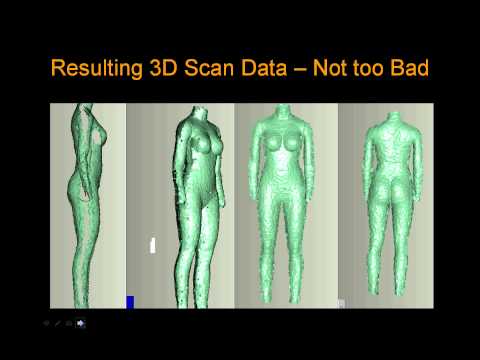
3D copies: 2013 Kinect scan (left figure) and peel 3d (right figure)
To improve the quality of scanning, the Canadian company USIMM has begun using peel 3d. The company specializes in the machining of non-metallic materials and is constantly involved in artistic projects. The main goal was to show the evolution of CNC machining by comparing the results of a 3D version of one employee made a few years ago with current results.
According to USIMM's Leia Lepage, scanning a living person is not an easy task: “Scanners are usually quite sensitive to the smallest movements, even if it's just breathing. Scanning a person is very difficult.” In order to accomplish such an extraordinary task, the USIMM team needed a 3D scanner that was immune to certain movements - but at the same time had high resolution and accuracy. Peel 3d possesses such qualities: a resolution of up to 0.5 mm and a volumetric accuracy of 0.5 mm/m.
The team scanned the same employee in the same pose as they did a few years ago. The resulting 3D scan data was sent to a five-axis CNC machine, and then a full-size replica of a person was created from polystyrene. The results were incredible.
The scan results obtained earlier by the Skanect device did not stand next to the peel 3d results. “The difference between the past and current results of the project is enormous,” commented Lea Lepage. The new copy turned out to be much more realistic and more accurately conveyed the original.
Materials provided by Creaform. Screensaver photo © Elisa Oddone, Al Jazeera
Article published on 12/08/2020, updated on 03/03/2022
3D scanning in medicine. Types of 3D scanners and applications in medicine
Contents
-
- Uses of 3D scanning in medicine
- Benefits of 3D scanning
- Types of 3D scanners
- Calibry
- Shining 3D EinScan H
- Predictions for the development of 3D technologies in medical practice
A 3D scanner is an essential technology in medical research centers that allows you to get an accurate model of the body or its individual parts. This is an essential attribute for diagnosing various diseases, visualizing the results of an operation or designing surgical prostheses.
Medical use of 3D scanning
3D scanning of a person is necessary for plastic surgeons, dentists, orthopedists and even cosmetologists. Before the advent of 3D technology, doctors had to take measurements manually, as well as create realistic dummies based on the information received: this is a hard and routine job that took a lot of time. 3D scanners have made medical procedures much easier: detailed information about a patient's anatomy can be obtained in minutes.
During the scan, the device recognizes the geometry of the body, and the sensors form a cloud of points. Based on the information received, the device software designs a polygonal body model and transfers it to the computer screen. The three-dimensional model becomes the basis for choosing the optimal method of surgical intervention, creating surgical prostheses, orthopedic shoes, and so on.
Benefits of 3D scanning
The 3D human scanner is used in all areas of medicine. Modern 3D scanners are replacing outdated diagnostic methods due to a number of advantages:
-
Three-dimensional models are more accurate and informative. At the diagnostic stage, some medical equipment makes errors, while 3D scanners show the best result;
-
Orthopedic corsets and insoles, created on the basis of a three-dimensional model of the patient, are much more accurate than plaster ones due to the consideration of aspects of individual anatomy;
-
Due to the high speed of work, new technologies allow you to get the necessary information urgently. Delay in medicine can cost a person's health and even life, and three-dimensional scanners help prevent the development of undesirable conditions;
-
Due to its safety, the technology is relevant for the diagnosis of adults and children.

Interesting! 3D scanning is used for operations on the cardiovascular system. Due to the high accuracy of the information received, surgeons choose the most relevant method of surgical intervention.
Types of 3D scanners
According to the principle of action in medicine, laser and optical 3D scanning is used. Each of these varieties has its own advantages, which we will discuss below.
Optical
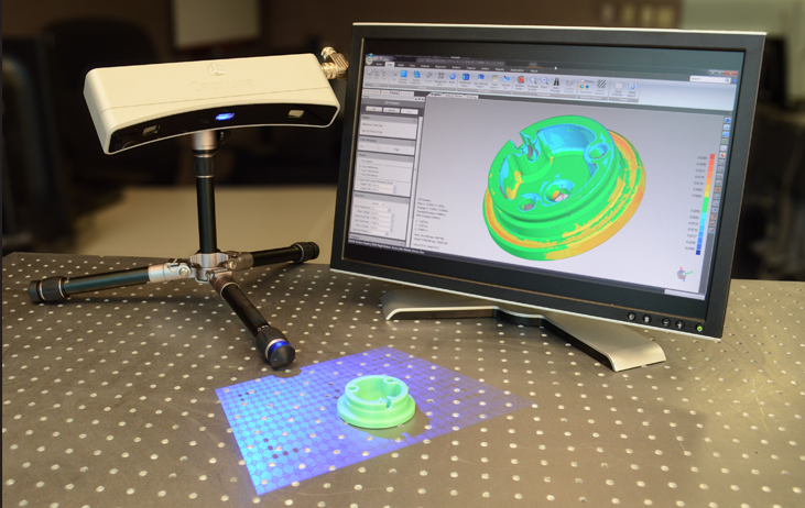
The areas to be examined are illuminated with structured light, while the cameras record the result from different angles. The area is illuminated with a strip of light or a specific pattern (black and white stripes or squares). The deformation of the light or pattern conveys to the device information about the shape and depth of the object. The process is recorded by one or two cameras, which transmit information about the structure of the object to the computer software.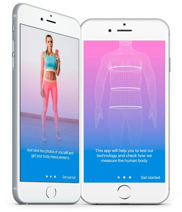
Such devices can be manual and desktop. In the first case, manual control of the device is used, in the second, the scanner is placed on the table. The main advantage of optical devices is the high speed of operation and the ability to transmit color.
Laser
The laser scanner measures the distance to the object under investigation and determines the actual length of the beams. The direction of the laser is controlled by encoders. Based on the analysis of the reflection of the rays, a cloud of points is formed, which makes up a three-dimensional image of the object under study. The main advantage of such devices is the ability to digitize objects with a complex shape.
Attention! For the most accurate scan results, the patient must remain static.
Add to compare
Product added to compare Go
| Manufacturer | Shining 3D |
Add to compare
Product added to compare Go
| Manufacturer | Shining 3D |
Add to compare
Product added to compare Go
| Manufacturer | Range Vision |
Add to compare
Product added to compare Go
| Manufacturer | Range Vision |
Main directions in medicine
Three-dimensional scanning is actively used in almost all medical fields.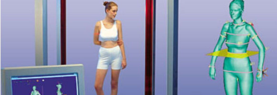 The presence of an accurate anatomical model helps to determine the current treatment regimen or to model a prosthesis. We must not forget the importance of virtual models for medical students: such schemes allow us to study the human anatomy in the most detailed way.
Dentistry
The use of an intraoral 3D scanner for diagnostics
The use of a dental 3D scanner to create a digital model of a plaster jaw prosthesis
Due to oncology, the Australian had to remove 80% of the upper jaw. The use of a 3D scanner helped create the perfect jaw prosthesis
3D dental scanning helps to create detailed virtual images of the patient's teeth. Thanks to this, the most complex restoration and restoration work is carried out, orthodontists design prostheses. The technology significantly reduces the time of manufacturing complex structures, and also saves the cost of producing plaster and other models.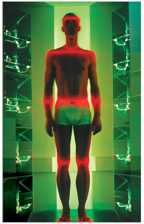
The 3D scanner is actively used in dentistry due to a number of advantages. The device is suitable for working with patients with an increased gag reflex. Just a few years ago, to create an impression of the teeth, an impression mass was placed in the patient's mouth: this procedure brought great discomfort. Nowadays, 3D-scanning of teeth is carried out contactlessly and does not bring discomfort.
Attention! The use of a 3D scanner in dental practice has a great advantage: the data obtained during the diagnostics are instantly transmitted to the laboratory.
Plastic surgery and cosmetology
face scan
Familiarization of the patient with the expected result of rhinoplasty based on a three-dimensional image of her face
3D face scanning is actively used in cosmetology and plastic surgery. The study is assigned to study problem areas before carrying out some cosmetic manipulations, as well as to design the expected result of plastic surgery.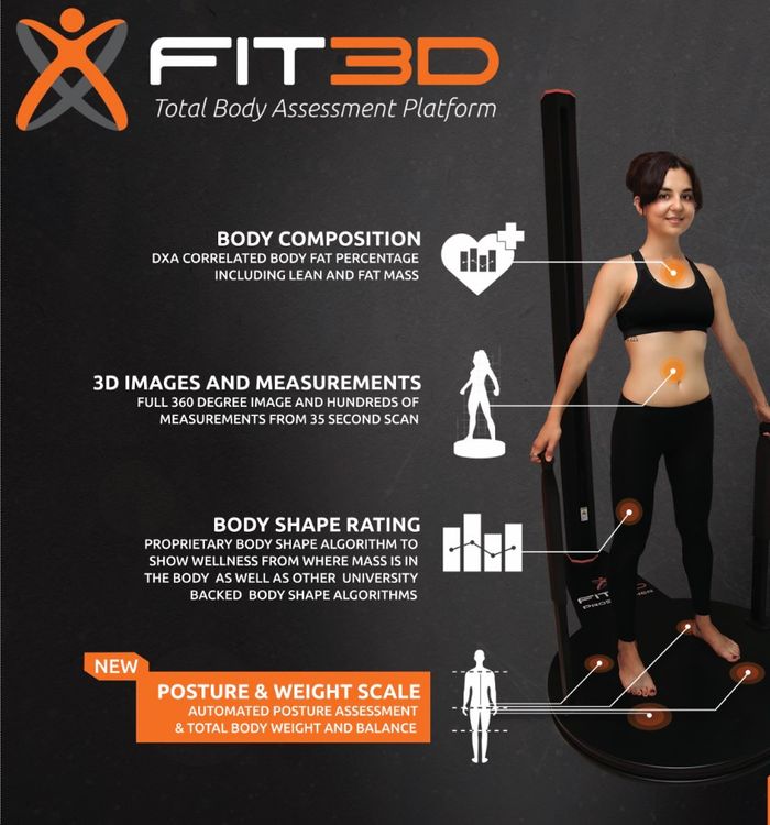 Thanks to this technology, the patient can get acquainted with how his face will look after surgery. The scanner is used for performing operations on the face (rhinoplasty, blepharoplasty) and body (mammoplasty, abdominoplasty, etc.).
Orthopedics
3D scanning of the spine
3D scanning of the foot to study the anatomical curves
Orthopedic insole printed on the basis of a 3D image of the foot
3D scanning of the body is relevant in the process of determining the type and stage of curvature of the spine or foot. The use of x-rays to diagnose the spine and lower extremities does not provide such accurate information, moreover, this procedure is contraindicated in some patients. 3D scanning helps to obtain detailed information about the pathology and develop actual orthopedic products (insoles and corsets).
For the manufacture of insoles, previously it was necessary to make an impression of the foot. If the doctor made mistakes, the cast turned out to be inaccurate, which affected the quality of the insoles. New technologies help to obtain the most accurate information, regardless of the doctor's actions.
Surgery
3D model of the heart created from scanned data
Siamese twin chest model. Based on the data obtained as a result of the scan, the surgeons managed to perform the most difficult operation to separate them.
The application of new technology plays an important role in surgery. Scanning is aimed at studying the features of the pathology, its location and blood supply. This information helps to carry out more accurate and safer operations, to prevent complications during surgery. In addition, scanning is used in the process of bioprinting tissues and organs for transplantation.
3D scanning is closely related to 3D printing: the surgeon gets the opportunity to study the model of the operated organ or part of the body, develop an actual scheme for surgical intervention and “hone” the movements.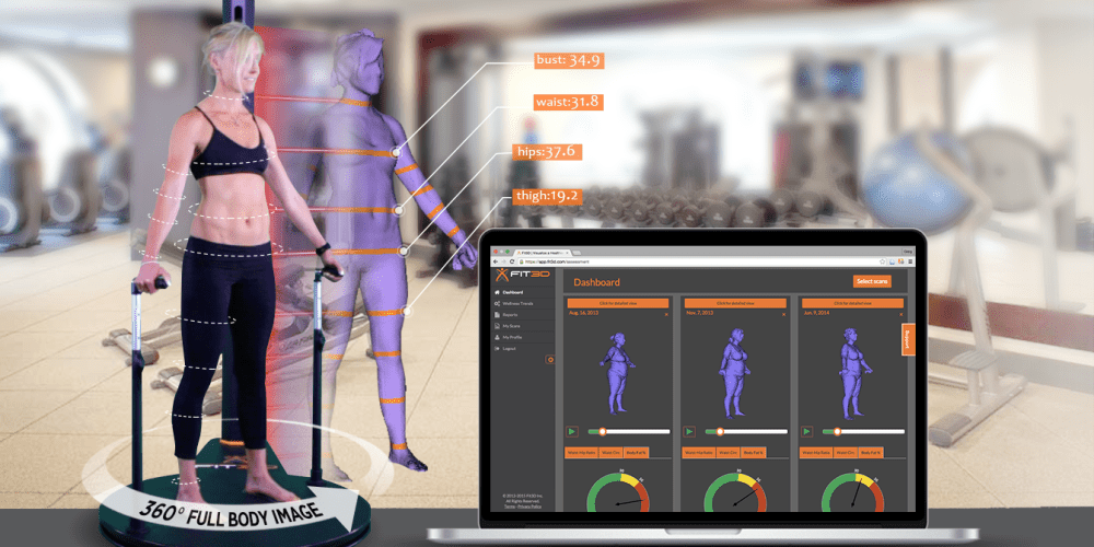
Prosthetics
The use of a 3D laser scanner in transplantology
Prosthesis based on scan data
In the process of creating manual prostheses, it is necessary to accurately determine the location of tissues, muscles and blood vessels. If errors are made during the diagnosis, local necrosis may develop due to tissue rejection. Modern technologies allow collecting the most accurate information about the shape of the stump and the location of tissues to create high-quality and comfortable prostheses.
Student education
A student studies pathological anatomy based on a 3D cadaver model
Scanning helps medical students study anatomy more effectively. The new method is much more efficient than the use of 2D images in classical anatomical atlases. Some programs allow you to observe the surgical intervention: for example, the trajectory of the scalpel during the operation.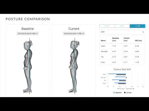
Interesting! In France, a 3D scanner is being used to study pathological anatomy. There was no need for an in-person autopsy, and the resulting schemes make it possible to study the human body in layers.
Add to compare
Product added to compare Go
| Manufacturer | Medit |
Add to compare
Product added to compare Go
| Manufacturer | 3Shape |
Add to compare
Product added to compare Go
| Manufacturer | ScanPod3D |
Add to compare
Product added to compare Go
| Manufacturer | Shining 3D |
Popular medical 3D scanners
A high-quality three-dimensional scanner provides accurate body diagnostics and successful operations. Next, we list a few scanners that have managed to establish themselves in the global market.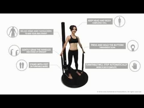
Shining Autoscan DS-EX
The device has an open, user-friendly platform that facilitates the scanning process. Various modules are easily installed on the platform, thanks to which the models are scanned in the articulator, the dental impression is examined from various angles, etc. A 3D scanner is used in a dental laboratory to create a virtual patient model, create a treatment plan, design crowns and jaw prostheses. The device is compact and highly accurate.
ScanPod 3D UPOD-S
An orthopedic scanner is used to obtain monochrome and color images. Despite its compactness, the device has a large scanning area, and high speed allows you to use the scanner in emergency mode. In addition, ScanPod 3D UPOD-S is capable of performing 40 operations on the resulting image, and also provides the ability to compare the anatomical features of the right and left foot. You can learn more about the features of the scanner in our review.
Medit i500
The intraoral 3D scanner allows you to scan the entire oral cavity in a matter of minutes. A 3D Full HD color image is displayed on the screen. A small scanning module provides convenience to the doctor during the examination and does not bring discomfort to the patient. Due to its low weight, it is convenient to use even during long-term diagnostics.
3Shape TRIOS 3 Basic Pod
The compact device provides maximum comfort for both doctor and patient during diagnostics. The intraoral scanner works non-contact: no special powders need to be used for the procedure. The resulting color image is sent to the computer screen in high resolution. The CAD/CAM software provides the ultimate in ease of use, and thanks to the USB port, the 3Shape TRIOS 3 Basic Pod can be connected to any computer.
ScanPod3D USOL
ScanPod3D USOL is a compact orthopedic scanner that allows you to quickly and accurately scan the foot of one foot.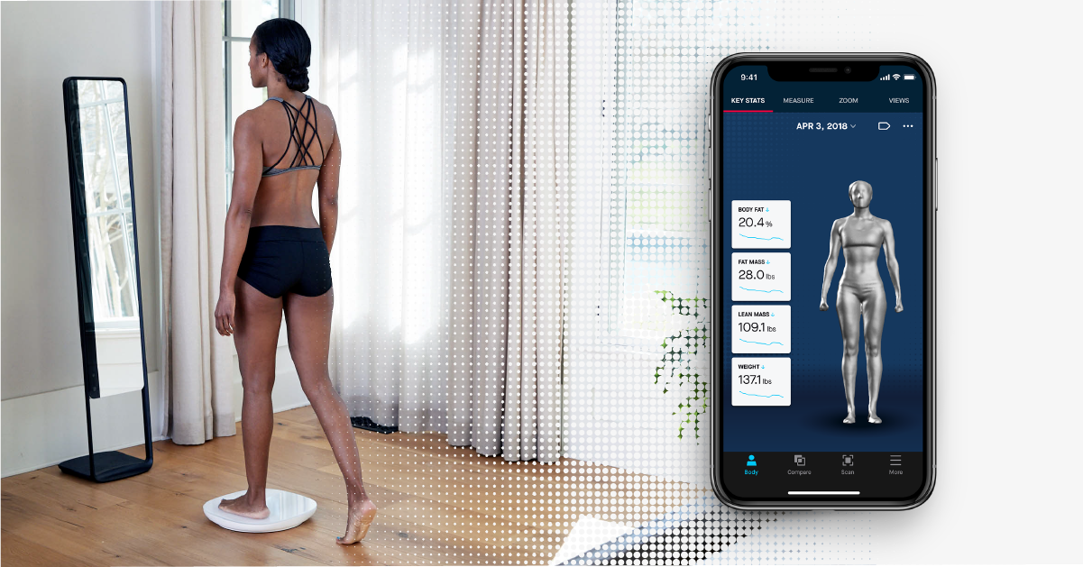 The resulting image can be scaled and rotated for detailed viewing. Diagnostics can be carried out without load, with partial and full load. The device scans the arch of the foot, insoles and lasts, and the RX shape setting is used to create orthopedic shoes.
Calibry
The portable handheld scanner allows you to get a digital image of objects from 20 cm to 10 m. Weight is 900 g, so operation is not difficult. The touch screen displays the result of the scan, so there is no need for constant checking with the computer. The scanner is used to examine the fine details, and the powerful software ensures easy operation. Calibry is used in 3D scanning of the spine, face, joints and other parts of the body.
Shining 3D EinScan H
The handheld scanner is lightweight and compact, making it easy to carry around. The use of the latest developments in data capture helped to achieve stunning scan results: up to 1,200,000 points per second.